Copyright
©The Author(s) 2023.
World J Clin Cases. Apr 16, 2023; 11(11): 2535-2540
Published online Apr 16, 2023. doi: 10.12998/wjcc.v11.i11.2535
Published online Apr 16, 2023. doi: 10.12998/wjcc.v11.i11.2535
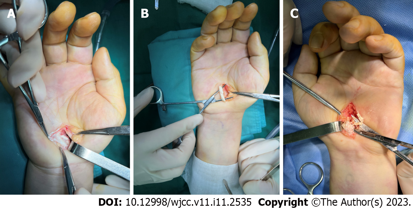
Figure 1 Intraoperative photographs of the surgical field of both hands.
A: The median nerve surrounded by miliary tophi in the synovium; B: The deep flexor tendon of the little finger of the right hand thickened by crystals deposited throughout the tendon; C: The deep flexor tendon of the ring finger of the left hand eroded by tophi.
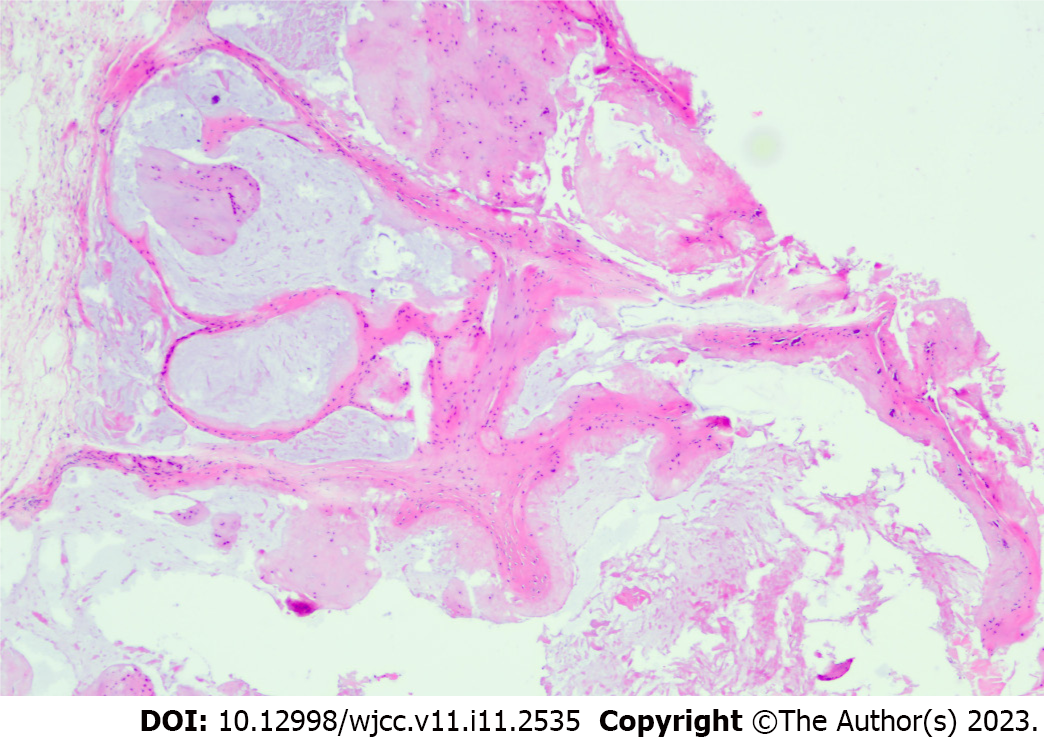
Figure 2 Histopathological findings.
Photomicrograph showing gout nodules filled with monosodium urate crystals (hematoxylin and eosin staining, magnification: eyepiece 10 × and objective 4 ×).
- Citation: Zhang GF, Rong CM, Li W, Wei BL, Han MT, Han QL. Bilateral carpal tunnel syndrome and motor dysfunction caused by gout and type 2 diabetes: A case report. World J Clin Cases 2023; 11(11): 2535-2540
- URL: https://www.wjgnet.com/2307-8960/full/v11/i11/2535.htm
- DOI: https://dx.doi.org/10.12998/wjcc.v11.i11.2535


