Copyright
©The Author(s) 2023.
World J Clin Cases. Apr 16, 2023; 11(11): 2489-2495
Published online Apr 16, 2023. doi: 10.12998/wjcc.v11.i11.2489
Published online Apr 16, 2023. doi: 10.12998/wjcc.v11.i11.2489
Figure 1 Image of an intracranial major artery embolism due to carotid artery thrombosis caused by a neck massager.
A: Computed tomography angiography (CTA) revealed occlusion of the M3 segment of the right middle cerebral artery; B: Magnetic resonance imaging performed 24 h after hospitalization revealed multiple acute infarcts in the cortex of the right cerebral hemisphere; C and D: CTA showed thrombus formation in the distal segment of the right and left sides of the common carotid arteries.
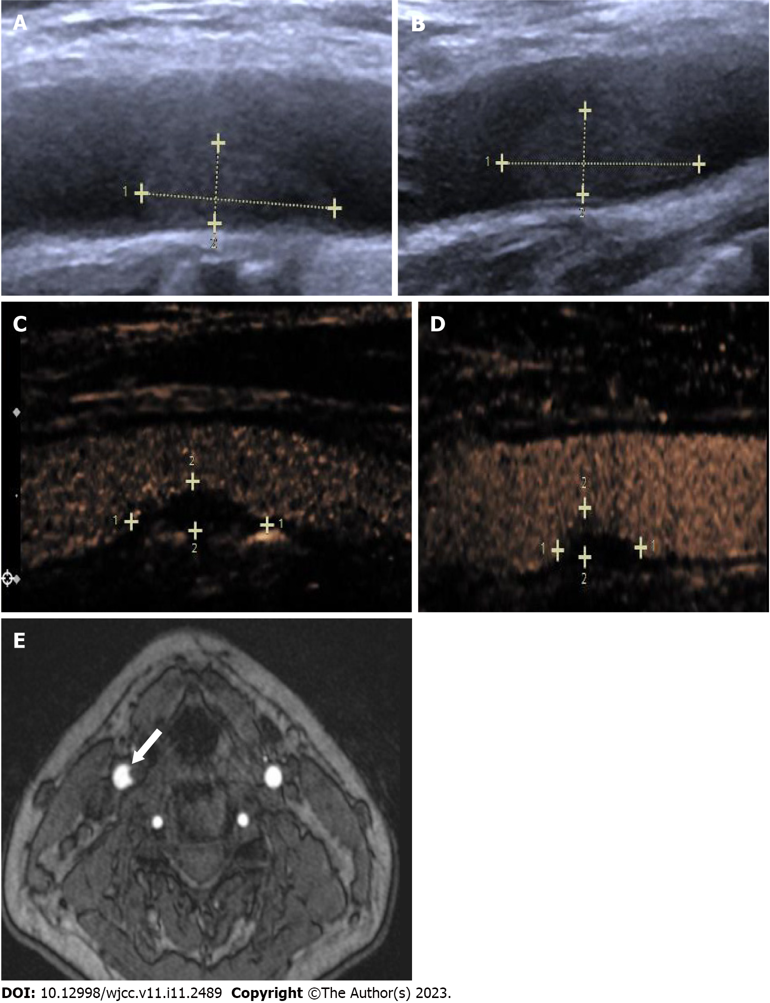
Figure 2 Carotid ultrasound and high-resolution enhanced magnetic resonance imaging of an intracranial major artery embolism due to carotid artery thrombosis caused by a neck massager.
A and B: Carotid ultrasound revealed that the right extent was approximately 12.8 mm × 3.8 mm, and the left extent was approximately 14.8 mm × 4.3 mm. Contrast-enhanced ultrasound showed that the thrombus was reduced on the fourth day, with no enhancement; C and D: The right extent was approximately 6.5 mm × 2.1 mm, and the left extent was approximately 6.4 mm × 2.4 mm; E: High-resolution enhanced magnetic resonance imaging of the neck vessels suggested a filling defect in the right common carotid arterie.
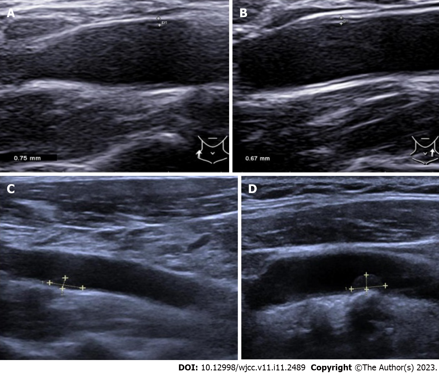
Figure 3 B-ultrasound of carotid thrombus changes after treatment.
A and B: On the eighth day, carotid ultrasound showed that the thrombus was further reduced, with an extent of approximately 6.5 mm × 2.4 mm on the right and 5.3 mm × 2.1 mm on the left; C and D: Carotid ultrasound examination performed on the 14th day revealed both right and left side common carotid arteries without thrombosis.
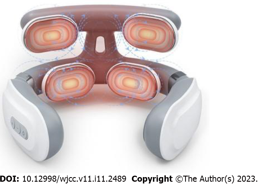
Figure 4 Structure of the neck massager used by the patient.
- Citation: Pan J, Wang JW, Cai XF, Lu KF, Wang ZZ, Guo SY. Intracranial large artery embolism due to carotid thrombosis caused by a neck massager: A case report. World J Clin Cases 2023; 11(11): 2489-2495
- URL: https://www.wjgnet.com/2307-8960/full/v11/i11/2489.htm
- DOI: https://dx.doi.org/10.12998/wjcc.v11.i11.2489


