Copyright
©The Author(s) 2023.
World J Clin Cases. Apr 6, 2023; 11(10): 2321-2328
Published online Apr 6, 2023. doi: 10.12998/wjcc.v11.i10.2321
Published online Apr 6, 2023. doi: 10.12998/wjcc.v11.i10.2321
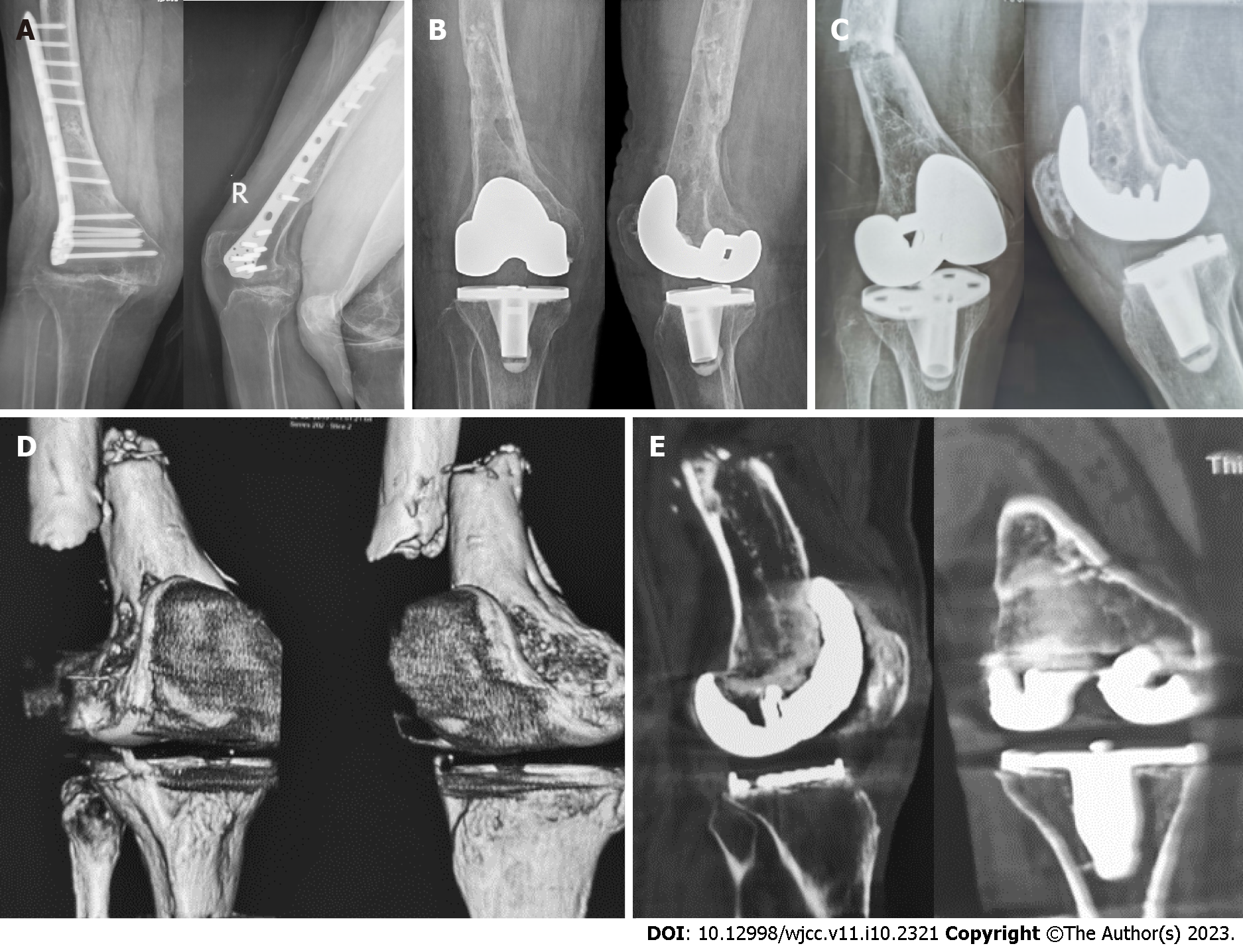
Figure 1 X-ray and computed tomography images.
A: Anterior–posterior and lateral X-ray images of the right knee before primary total knee arthroplasty showing an end-stage osteoarthritis of the right knee with valgus deformity and a hardware fixed on right femur without signs of fracture (December 2018); B: Anterior–posterior and lateral X-ray images of the right knee after the primary total knee arthroplasty showing satisfactory position of the knee prosthesis and that the original hardware was completely removed without fracture (December 2018); C: Anterior–posterior and lateral X-ray images of the right knee after fall showing the distal femur fracture with significant displacement and that the knee prosthesis was seemingly stable (July 2019); D: Three-dimensional computed tomography (CT) images showing fractures of the right distal femur with severe displacement (July 2019); E: Coronal and sagittal CT images showing radiolucent lines around the femoral and tibial prosthesis (July 2019).
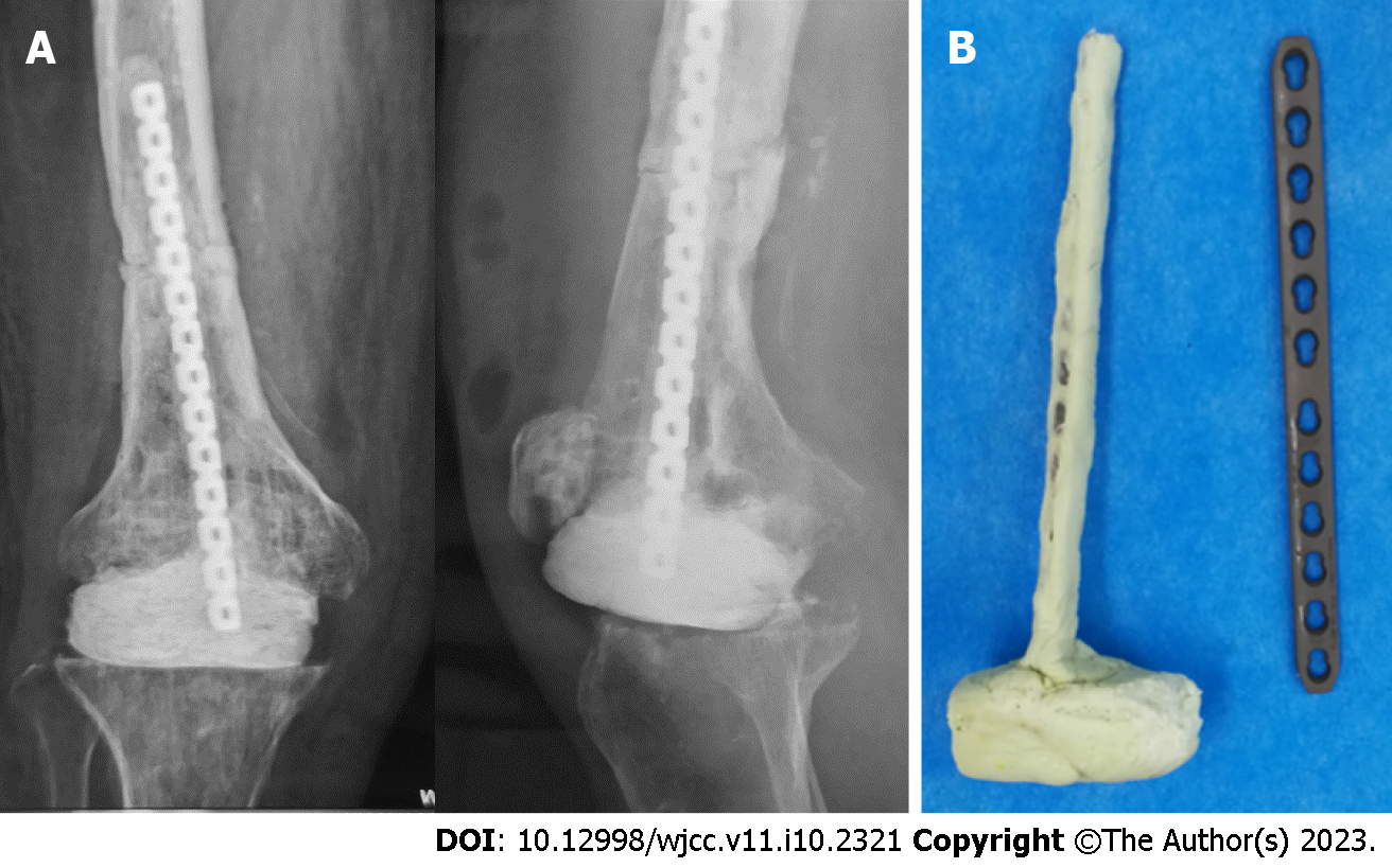
Figure 2 X-ray and intraoperative images.
A: Anterior–posterior and lateral X-ray images of the right knee after the first stage operation showing favorable position of the cement spacer and satisfactory reduction and fixation of the femoral fracture (July 2019); B: A hand-made T-shaped antibiotic-laden cement spacer for knee gap occupation and femoral fracture fixation.
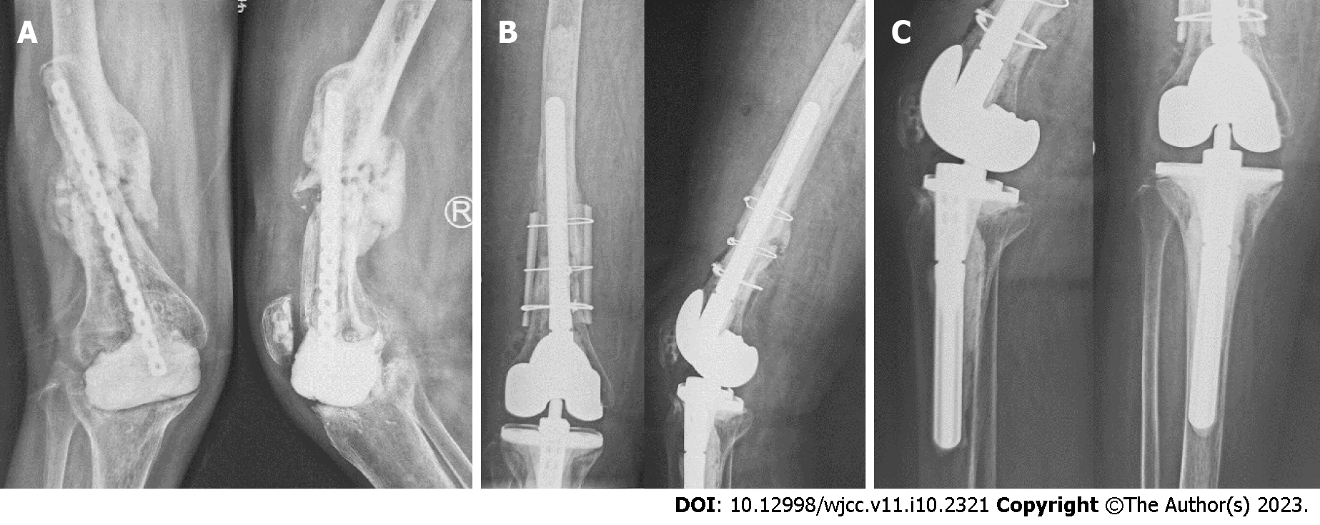
Figure 3 X-ray images.
A: Anterior–posterior and lateral X-ray images of the right knee before the second stage operation showing that the cement spacer slightly displaced to the medial side, the proximal end of internal fixator came out through the middle femoral shaft, callus formed at the previous fracture of the femur but not healing yet, and bone defects existed in the distal femur and proximal tibia, which resulted in valgus deformity of the right knee (May 2020); B and C: Anterior–posterior and lateral X-ray images of the right knee after the second stage operation showing no loosening of cemented revision knee prosthesis and that the femoral fracture was fixed with intramedullary stem and extramedullary cortical splints (May 2020).
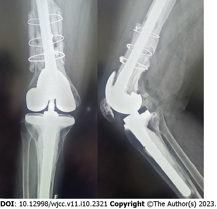
Figure 4 X-ray images.
Anterior–posterior and lateral X-ray images showing a large amount of new bone formed between the cortical splits and femoral shaft with no loosening of the knee prosthesis (August 2022).
- Citation: Hao LJ, Wen PF, Zhang YM, Song W, Chen J, Ma T. Treatment of periprosthetic knee infection and coexistent periprosthetic fracture: A case report and literature review. World J Clin Cases 2023; 11(10): 2321-2328
- URL: https://www.wjgnet.com/2307-8960/full/v11/i10/2321.htm
- DOI: https://dx.doi.org/10.12998/wjcc.v11.i10.2321


