Copyright
©The Author(s) 2023.
World J Clin Cases. Apr 6, 2023; 11(10): 2301-2307
Published online Apr 6, 2023. doi: 10.12998/wjcc.v11.i10.2301
Published online Apr 6, 2023. doi: 10.12998/wjcc.v11.i10.2301
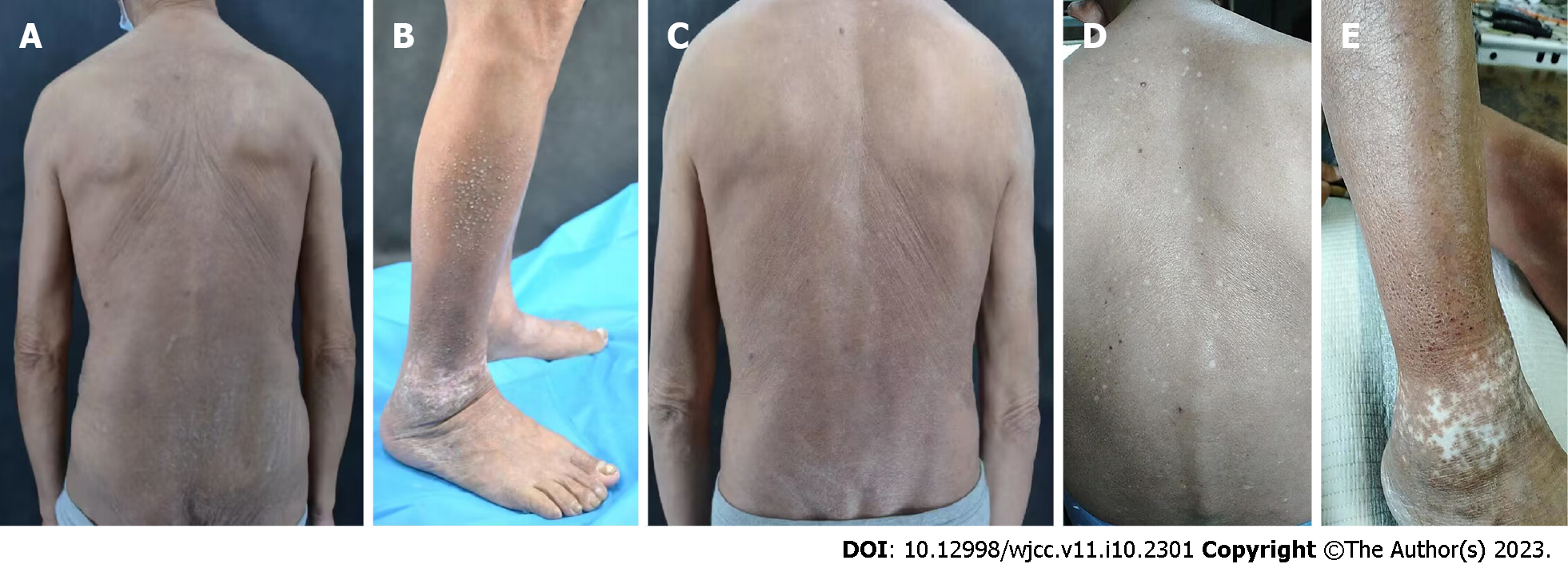
Figure 1 Skin lesions at various follow-up time points of patient 1 after the application of dupilumab to the lesions.
A: Skin lesions on the back before the treatment of dupilumab; B: Skin lesions on both lower limbs before the treatment of dupilumab; C: Posterior trunk at 16-wk follow-up; D: Back at 55-wk follow-up; E: Skin lesions on both lower limbs at 55-wk follow-up.
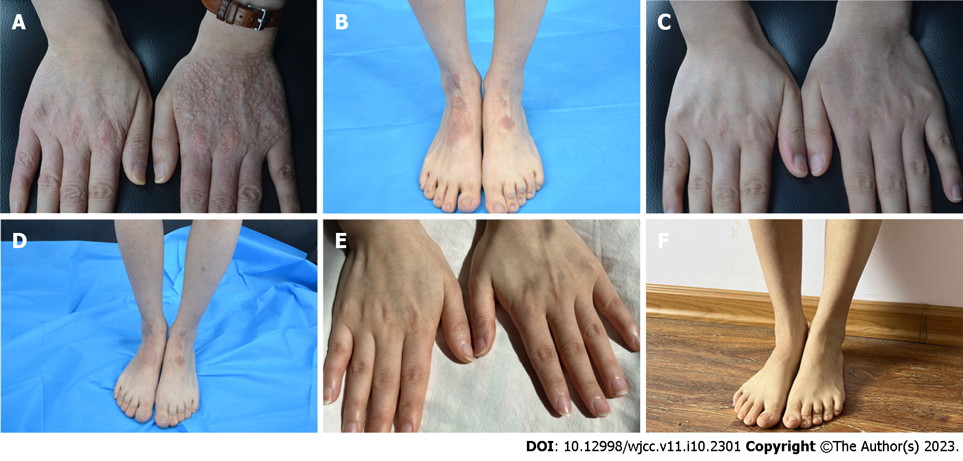
Figure 2 Skin lesions at various follow-up time points of patient 2 after application of dupilumab to the lesions.
A: Skin lesions of hands before the treatment of dupilumab; B: Skin lesions of both lower limbs before the treatment of dupilumab; C: 16-wk follow-up; D: 16-wk follow-up; E: Hands 1.5 year post-treatment follow-up; F: Lower limbs 1.5 years post-treatment follow-up.
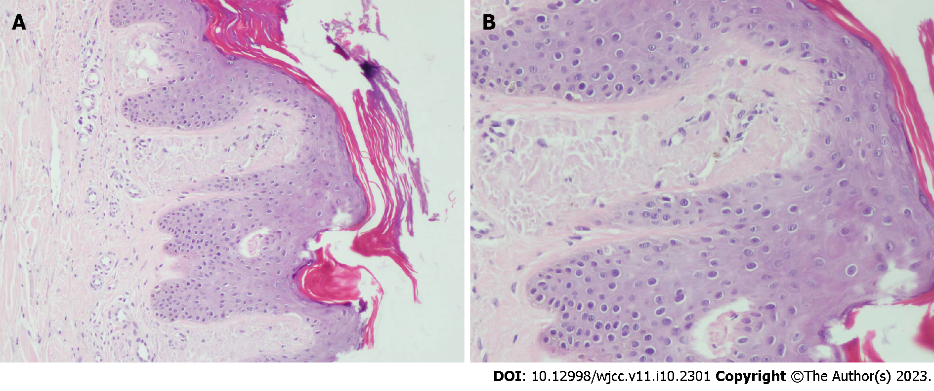
Figure 3 Skin histopathology.
A: Amorphous amyloid lump deposition with mild eosinophilic staining in the dermal papillary layer, consistent with amyloidosis (hematoxylin-eosin staining, 100 ×); B: Amorphous amyloid lump deposition with mild eosinophilic staining in the dermal papillary layer, consistent with amyloidosis (hematoxylin-eosin staining, 400 ×).
- Citation: Zhao XQ, Zhu WJ, Mou Y, Xu M, Xia JX. Dupilumab for treatment of severe atopic dermatitis accompanied by lichenoid amyloidosis in adults: Two case reports. World J Clin Cases 2023; 11(10): 2301-2307
- URL: https://www.wjgnet.com/2307-8960/full/v11/i10/2301.htm
- DOI: https://dx.doi.org/10.12998/wjcc.v11.i10.2301


