Copyright
©The Author(s) 2023.
World J Clin Cases. Apr 6, 2023; 11(10): 2276-2281
Published online Apr 6, 2023. doi: 10.12998/wjcc.v11.i10.2276
Published online Apr 6, 2023. doi: 10.12998/wjcc.v11.i10.2276
Figure 1
Pathological result suggestive of a paraganglioma (HE × 400).
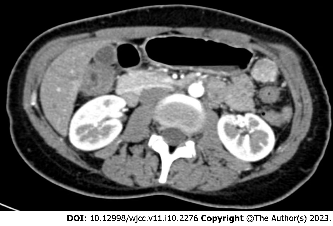
Figure 2
Contrast-enhanced computed tomography showing a mass in the left upper abdomen, indicating either a benign mesenchymal tumor or an ectopic accessory spleen.
- Citation: Guo W, Li WW, Chen MJ, Hu LY, Wang XG. Primary intra-abdominal paraganglioma: A case report. World J Clin Cases 2023; 11(10): 2276-2281
- URL: https://www.wjgnet.com/2307-8960/full/v11/i10/2276.htm
- DOI: https://dx.doi.org/10.12998/wjcc.v11.i10.2276


