Copyright
©The Author(s) 2023.
World J Clin Cases. Apr 6, 2023; 11(10): 2201-2212
Published online Apr 6, 2023. doi: 10.12998/wjcc.v11.i10.2201
Published online Apr 6, 2023. doi: 10.12998/wjcc.v11.i10.2201
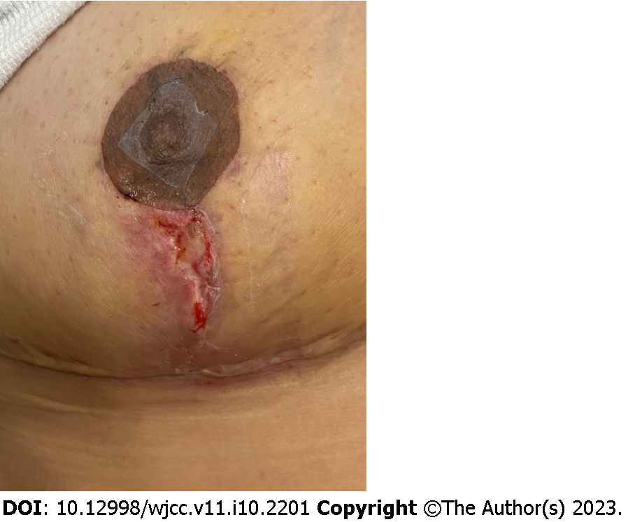
Figure 1 Wound dehiscence after implant-based breast reconstruction.
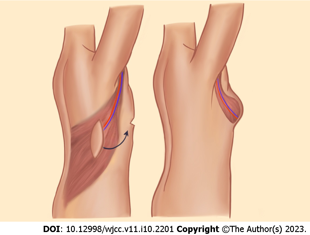
Figure 2 Latissimus dorsi myocutaneous flap based on the thoracodorsal vessels.
Figure 3 Pedicled transverse rectus abdominis myocutaneous flap uses lower abdominal adipose tissue and is based on the superior epigastric artery.
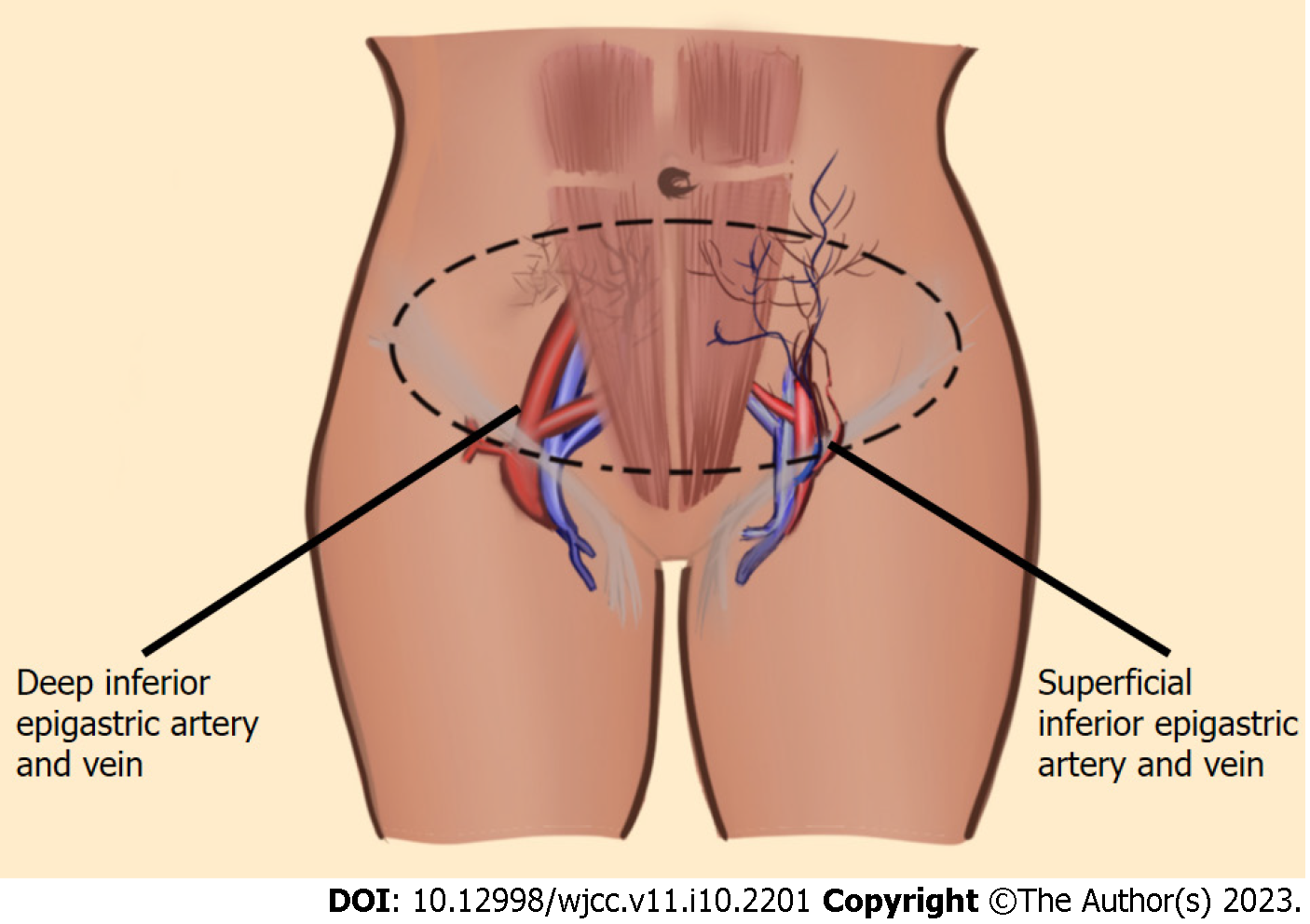
Figure 4 Abdomen-based free flaps are based on superficial or deep inferior epigastric vessels.
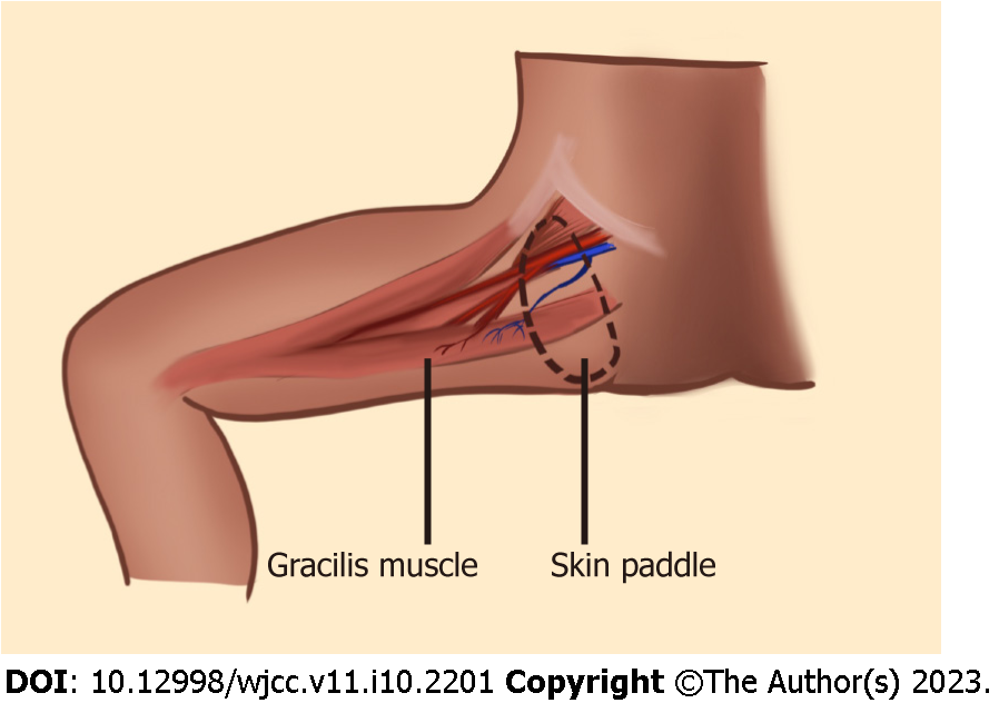
Figure 5 Transverse upper gracilis flap is based on the medial circumflex femoral artery which is a branch of the profunda femoris artery.
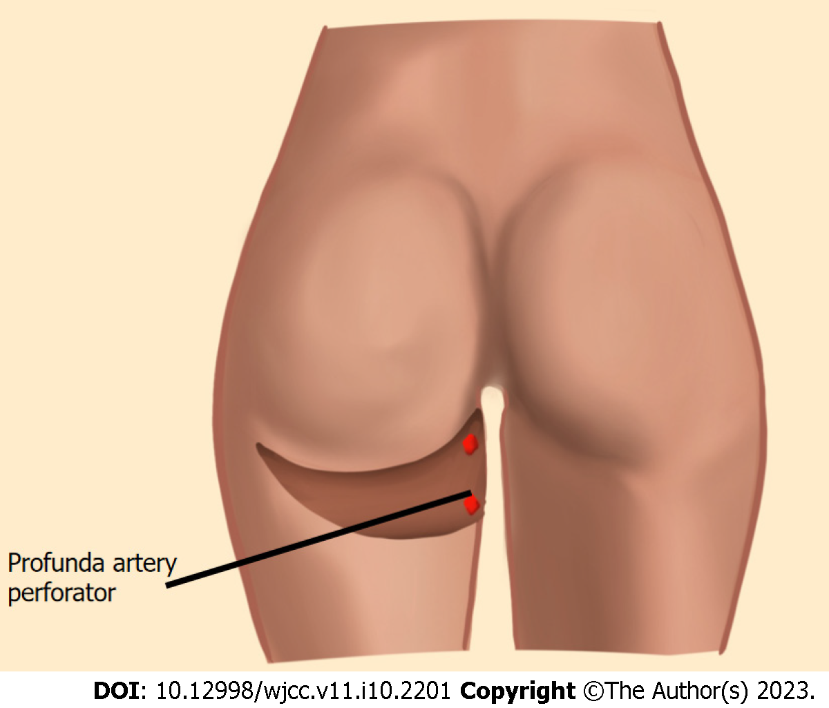
Figure 6 Profunda artery perforator flap carries posterior thigh skin and adipose tissue.
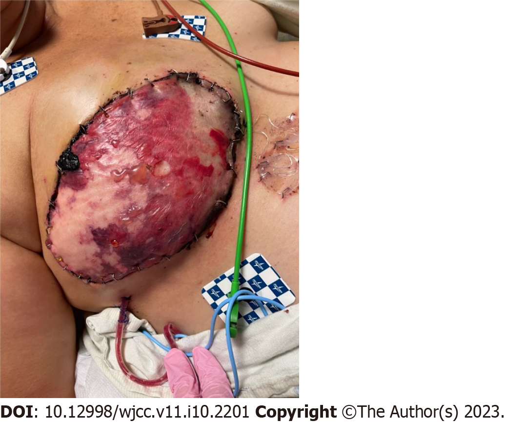
Figure 7 Partial flap loss with free deep inferior epigastric perforator flap in evolution.
Figure 8 Infected pedicled latissimus dorsi flap with implant.
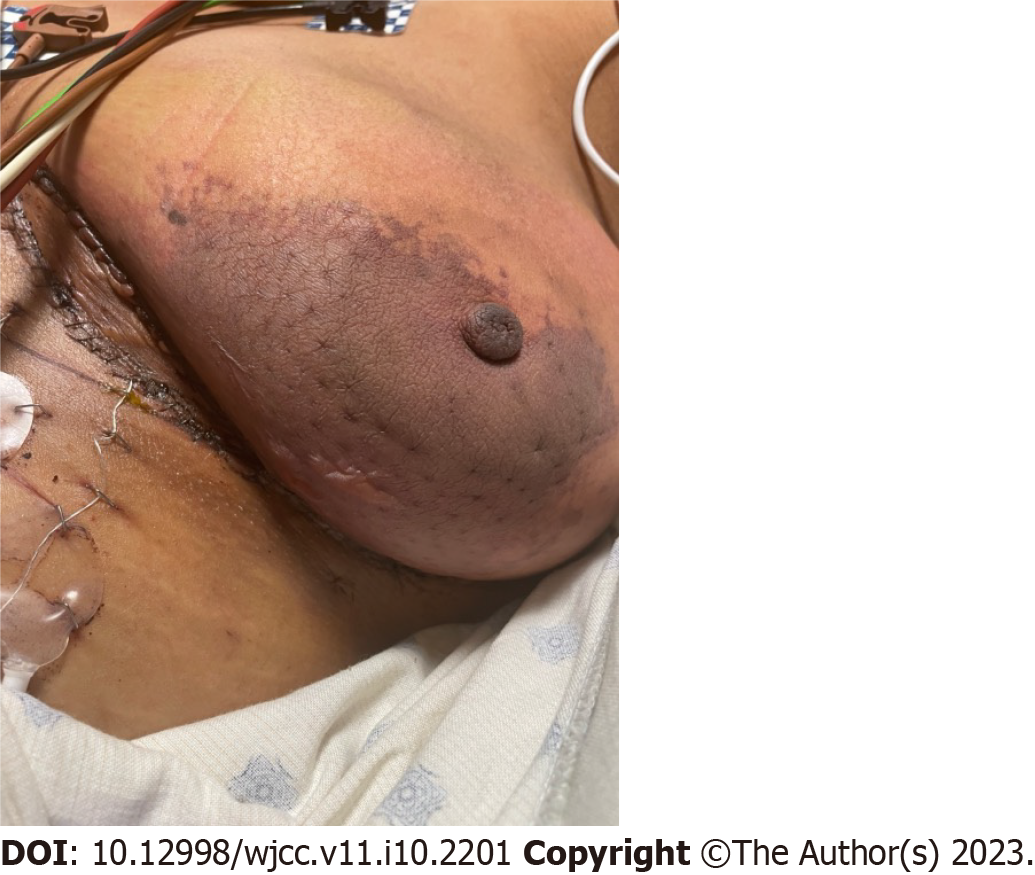
Figure 9 Hematoma of the mastectomy flap following deep inferior epigastric perforator flap breast reconstruction.
- Citation: Malekpour M, Malekpour F, Wang HTH. Breast reconstruction: Review of current autologous and implant-based techniques and long-term oncologic outcome. World J Clin Cases 2023; 11(10): 2201-2212
- URL: https://www.wjgnet.com/2307-8960/full/v11/i10/2201.htm
- DOI: https://dx.doi.org/10.12998/wjcc.v11.i10.2201


