Copyright
©The Author(s) 2023.
World J Clin Cases. Apr 6, 2023; 11(10): 2181-2188
Published online Apr 6, 2023. doi: 10.12998/wjcc.v11.i10.2181
Published online Apr 6, 2023. doi: 10.12998/wjcc.v11.i10.2181
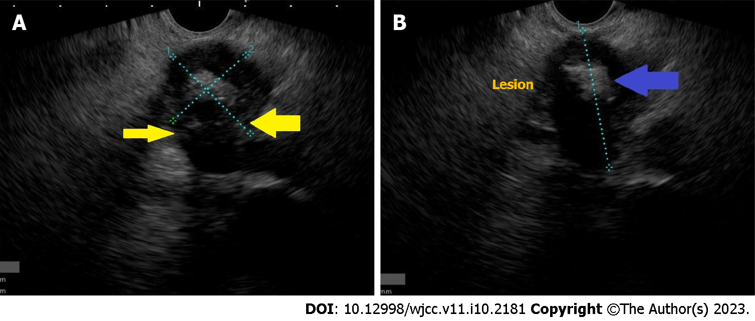
Figure 1 Endoscopic ultrasound of pancreatic tuberculosis.
A: Image showing a pancreatic lesion respecting vessels (yellow arrow), B: Pancreatic lesion with central calcification (blue arrow).
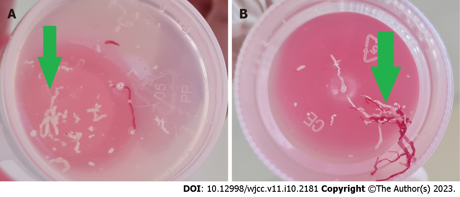
Figure 2 Cheesy macroscopic on-site evaluation (green arrow).
A: The core of the first two passes was cheesy; B: The third one was also cheesy but a little bloody.
- Citation: Delsa H, Bellahammou K, Okasha HH, Ghalim F. Cheesy material on macroscopic on-site evaluation after endoscopic ultrasound-guided fine-needle biopsy: Don't miss the tuberculosis. World J Clin Cases 2023; 11(10): 2181-2188
- URL: https://www.wjgnet.com/2307-8960/full/v11/i10/2181.htm
- DOI: https://dx.doi.org/10.12998/wjcc.v11.i10.2181


