Copyright
©The Author(s) 2022.
World J Clin Cases. Mar 26, 2022; 10(9): 2764-2772
Published online Mar 26, 2022. doi: 10.12998/wjcc.v10.i9.2764
Published online Mar 26, 2022. doi: 10.12998/wjcc.v10.i9.2764
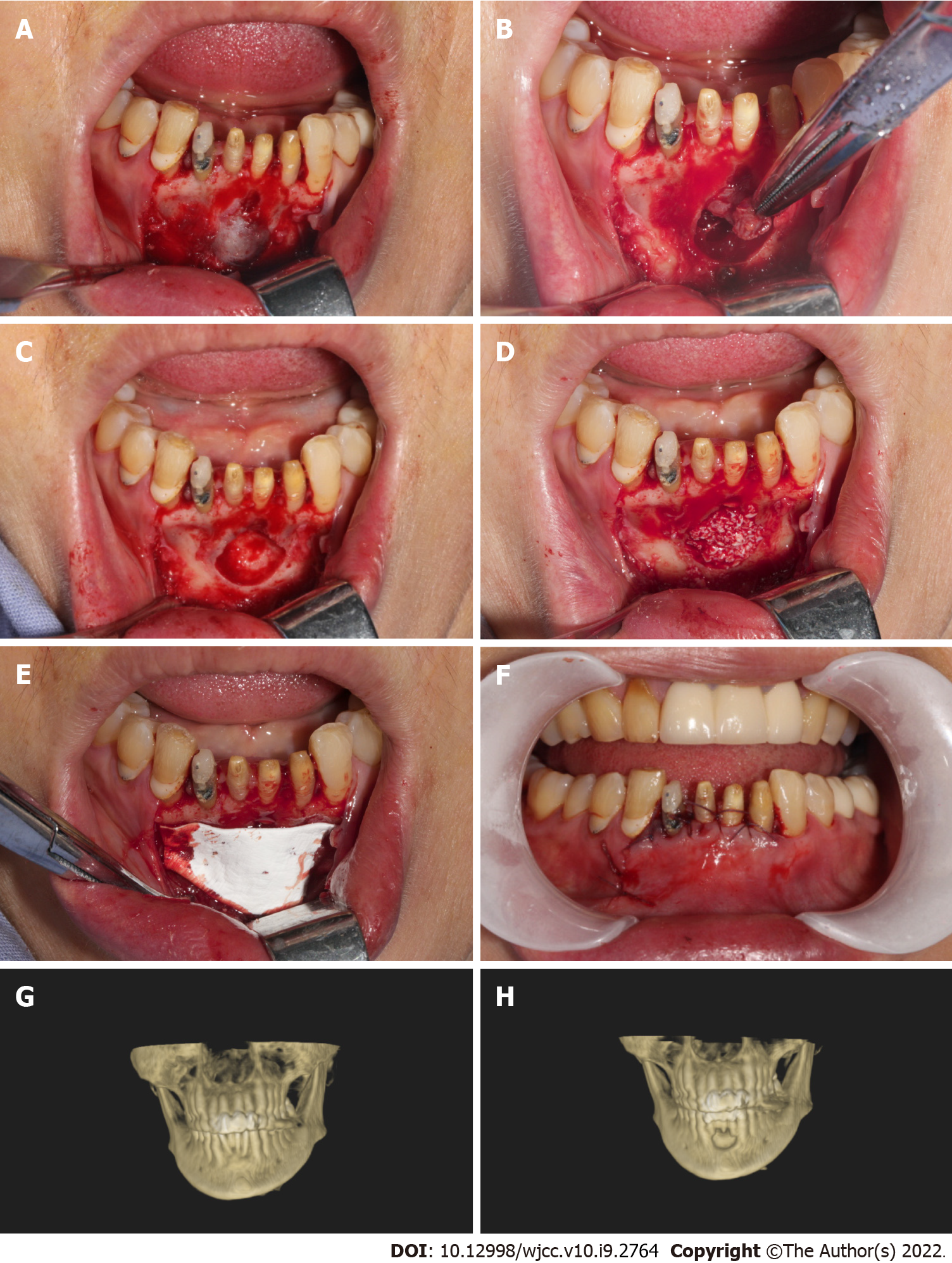
Figure 1 Photographic observation of cystic cavity.
A: Trapezoidal flap gingival sulcus incision, exposed bone surface; B: Cyst enucleation; C: Bone cavity; D: Implanted bone powder (Bio-Oss, Geistlich Pharma, Princeton, NJ, United States); E: Overed by a resorbable collagen membrane (Bio-Gide, Geistlich Pharma, Princeton, NJ, United States); F: Suture; G: Preoperative; H: Postoperative.
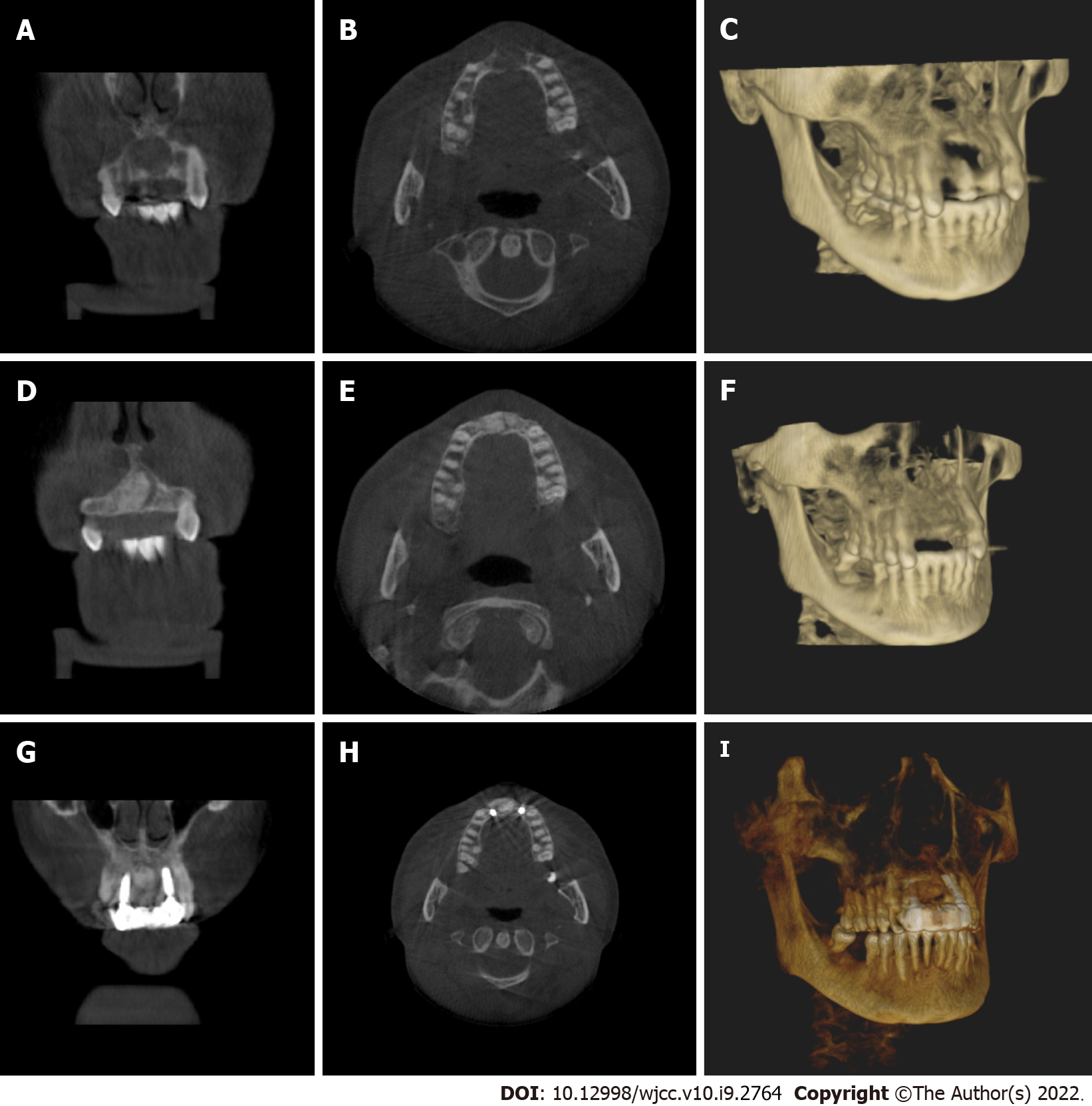
Figure 2 The patients with tooth defects were treated with implants.
A-C: Preoperative imaging; D-F: The imaging of six month after operation; G, H, and I: The imaging of nine month after operation, implant surgery; A, D, and G: Coronal plane; B, E, and H: Horizontal plane; C, F, and I: Three-dimensional reconstruction.
Figure 3 Clinical observation of postoperative treatment effect.
A: Six months after cyst enucleation combined with guided bone regeneration (GBR); B-D: Nine months after cyst enucleation combined with GBR, implant surgery and immediate restoration; E and F: Final implant-supported prosthesis.
- Citation: Cao YT, Gu QH, Wang YW, Jiang Q. Enucleation combined with guided bone regeneration in small and medium-sized odontogenic jaw cysts . World J Clin Cases 2022; 10(9): 2764-2772
- URL: https://www.wjgnet.com/2307-8960/full/v10/i9/2764.htm
- DOI: https://dx.doi.org/10.12998/wjcc.v10.i9.2764


