Copyright
©The Author(s) 2022.
World J Clin Cases. Mar 16, 2022; 10(8): 2637-2643
Published online Mar 16, 2022. doi: 10.12998/wjcc.v10.i8.2637
Published online Mar 16, 2022. doi: 10.12998/wjcc.v10.i8.2637
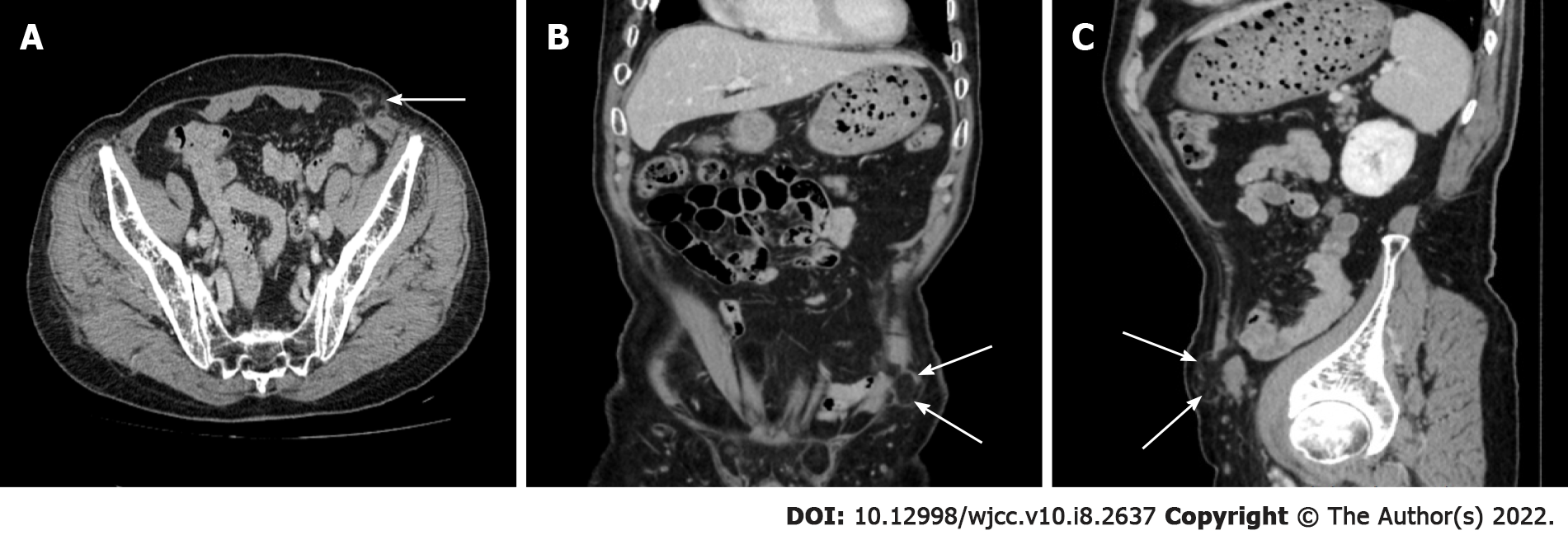
Figure 1 Preoperative computed tomography images of an abdominal wall hernia at the former drain-site in the left lower flank.
A: Transverse section; B: Coronal section; C: Sagittal section. The protruded content was confirmed to be the large omentum (white arrow).
- Citation: Su J, Deng C, Yin HM. Drain-site hernia after laparoscopic rectal resection: A case report and review of literature. World J Clin Cases 2022; 10(8): 2637-2643
- URL: https://www.wjgnet.com/2307-8960/full/v10/i8/2637.htm
- DOI: https://dx.doi.org/10.12998/wjcc.v10.i8.2637


