Copyright
©The Author(s) 2022.
World J Clin Cases. Mar 16, 2022; 10(8): 2584-2590
Published online Mar 16, 2022. doi: 10.12998/wjcc.v10.i8.2584
Published online Mar 16, 2022. doi: 10.12998/wjcc.v10.i8.2584
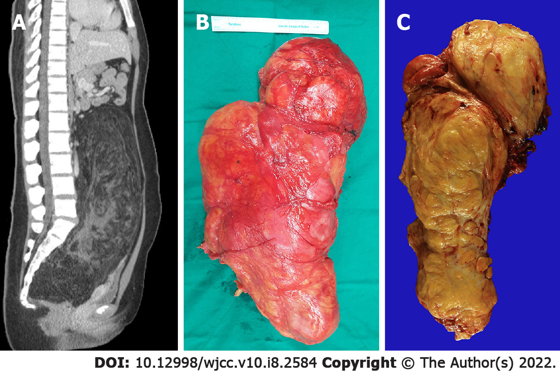
Figure 1 Abdominal computed tomographic findings and gross findings.
A: Large fatty mass with a complex soft tissue component. The tumor was filled the entire abdominal cavity and pushed up the abdominal viscera from the pelvic cavity; B: The tumor weigh approximately 5000 g, had an egg-shaped appearance, and mesures 38 cm × 24 cm × 11.5 cm in size; C: The cut surface show a large fatty mass with diffuse heterogeneous fibrotic change. No hemorrhage or necrosis is observed.
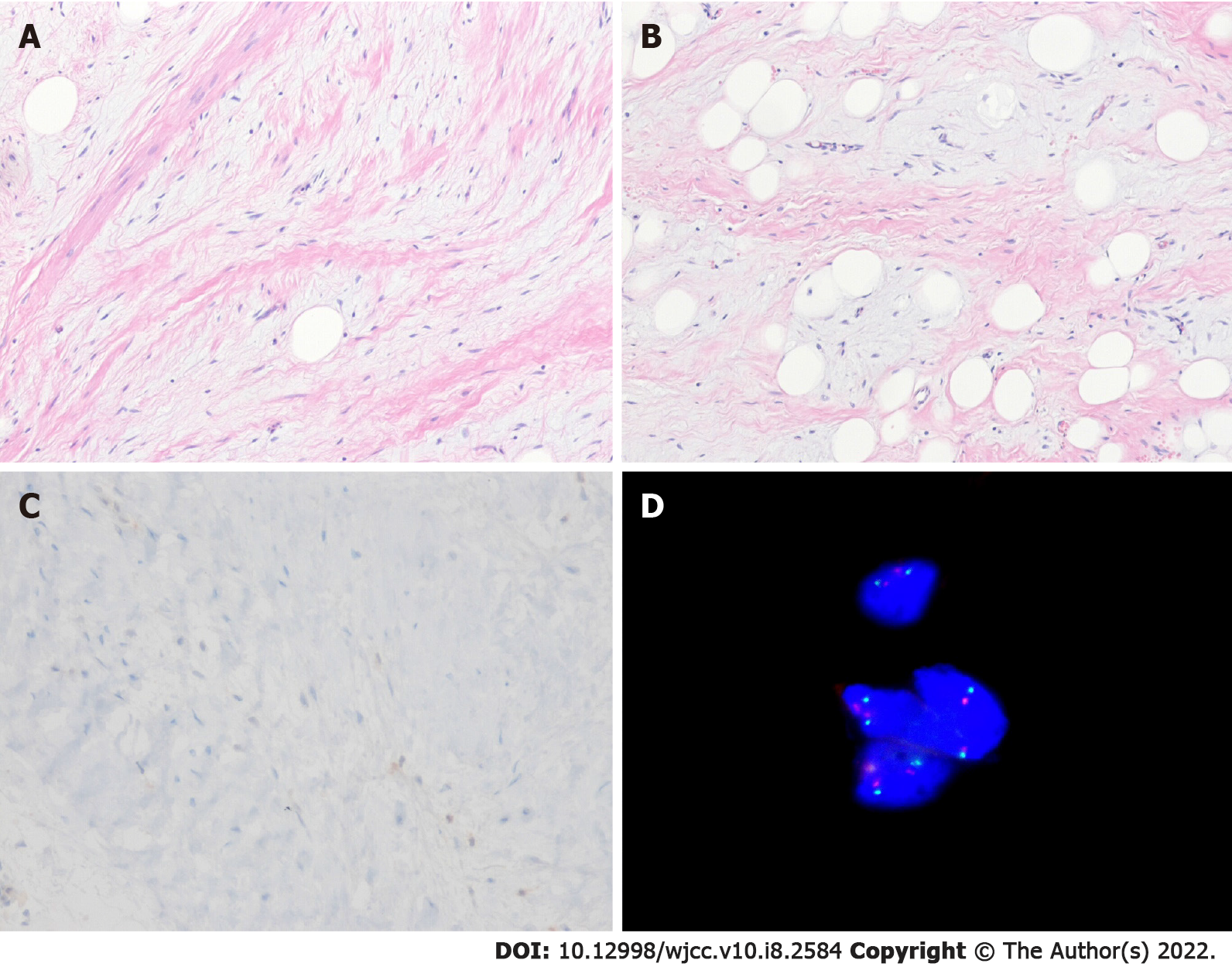
Figure 2 Microscopic findings.
A: The neoplasm shows spindle cells with mild nuclear atypia and hyperchromatia. There are admixed adipocytes (Hematoxylin and eosin stain, × 100); B: The variation of adipocytic size and myxoid stroma are present (Hematoxylin and eosin stain, × 100); C: The spindle cells are negative for MDM2 (Immunohistochemical stain, × 200); D: Fluorescence in situ hybridization shows no MDM2 amplification.
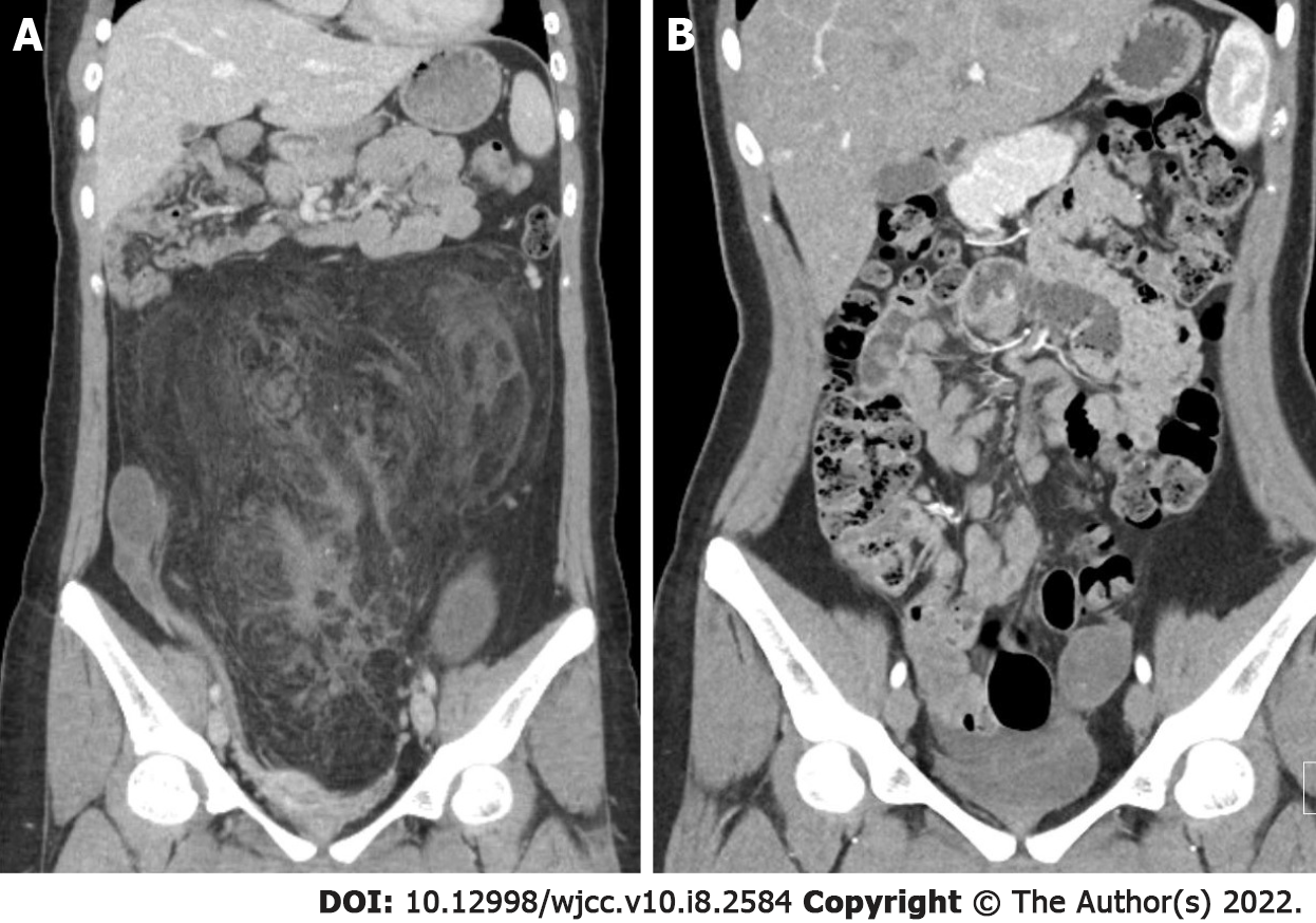
Figure 3 Computed tomography.
A: Pre-operative computed tomography findings; B: post-operative follow up computed tomography findings. At the 12-mo follow-up, no evidence of recurrence or metastasis was observed.
- Citation: Bae JM, Jung CY, Yun WS, Choi JH. Large retroperitoneal atypical spindle cell lipomatous tumor, an extremely rare neoplasm: A case report. World J Clin Cases 2022; 10(8): 2584-2590
- URL: https://www.wjgnet.com/2307-8960/full/v10/i8/2584.htm
- DOI: https://dx.doi.org/10.12998/wjcc.v10.i8.2584


