Copyright
©The Author(s) 2022.
World J Clin Cases. Mar 16, 2022; 10(8): 2569-2576
Published online Mar 16, 2022. doi: 10.12998/wjcc.v10.i8.2569
Published online Mar 16, 2022. doi: 10.12998/wjcc.v10.i8.2569
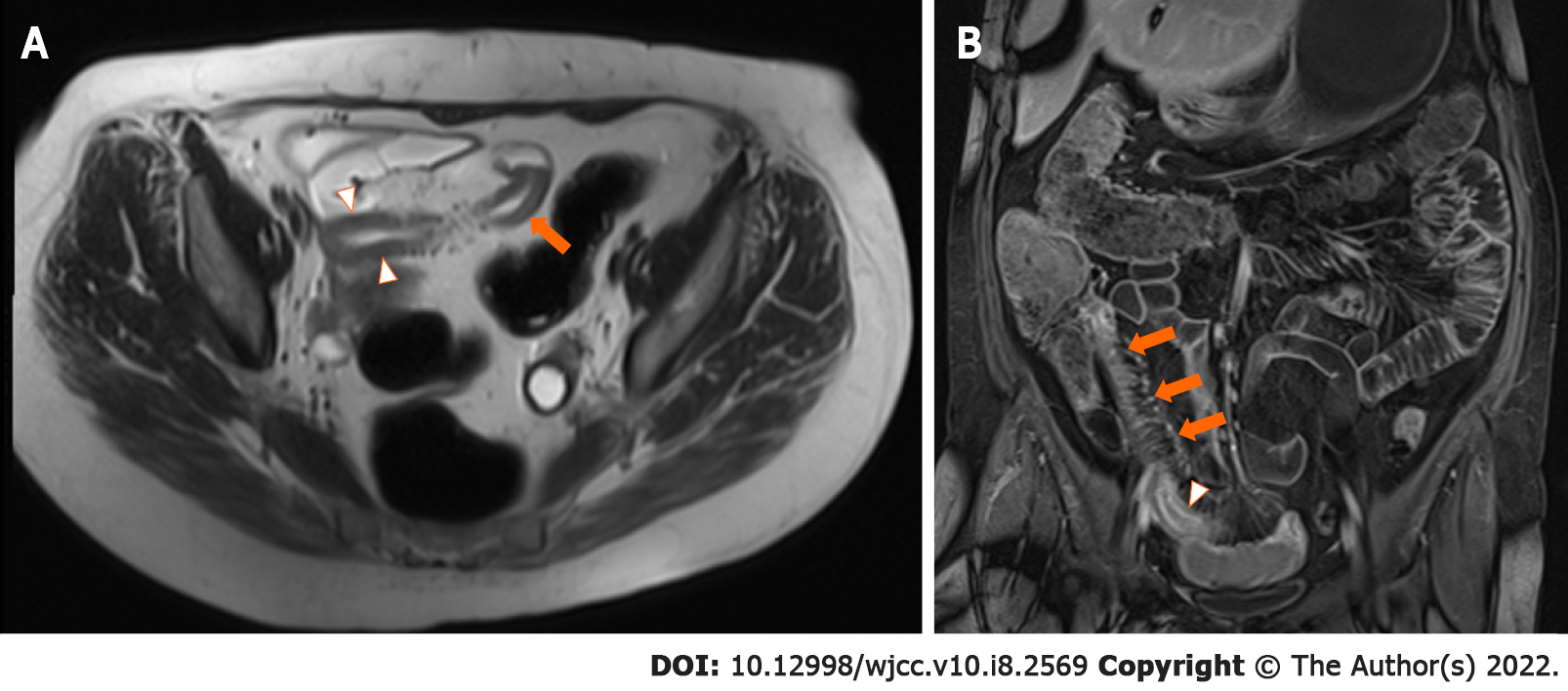
Figure 1 Repeat magnetic resonance enterography (original images).
A: Axial T2 image demonstrating bowel wall thickening (arrowheads) and T2 hyperintensity within the bowel wall (arrow), consistent with mural oedema; B: Coronal contrast-enhanced T1 fat-suppressed image demonstrating mural hyperenhancement (arrowhead) and extensive mesenteric vascular congestion, known as the ‘comb sign’ (arrows).
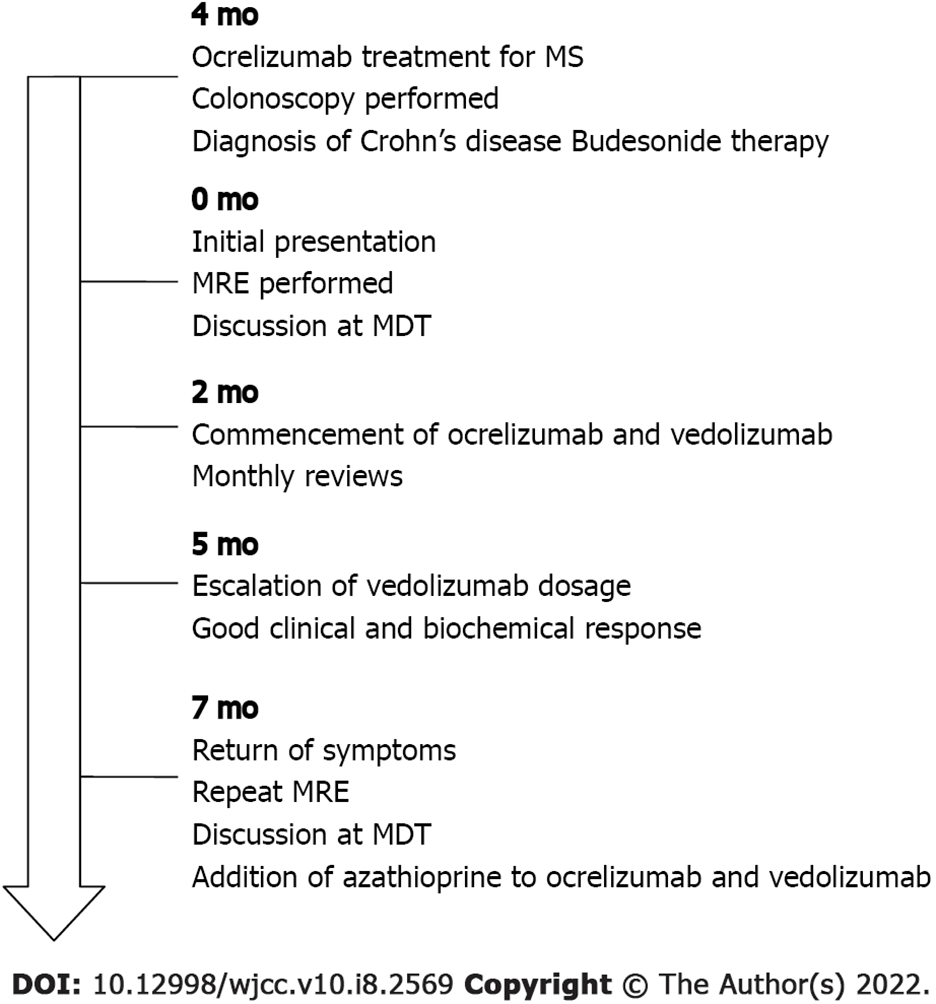
Figure 2 Timeline of events.
MS: Multiple sclerosis; MDT: Multidisciplinary Team; MRE: Magnetic resonance enterography.
- Citation: Au M, Mitrev N, Leong RW, Kariyawasam V. Dual biologic therapy with ocrelizumab for multiple sclerosis and vedolizumab for Crohn’s disease: A case report and review of literature. World J Clin Cases 2022; 10(8): 2569-2576
- URL: https://www.wjgnet.com/2307-8960/full/v10/i8/2569.htm
- DOI: https://dx.doi.org/10.12998/wjcc.v10.i8.2569


