Copyright
©The Author(s) 2022.
World J Clin Cases. Mar 16, 2022; 10(8): 2529-2536
Published online Mar 16, 2022. doi: 10.12998/wjcc.v10.i8.2529
Published online Mar 16, 2022. doi: 10.12998/wjcc.v10.i8.2529
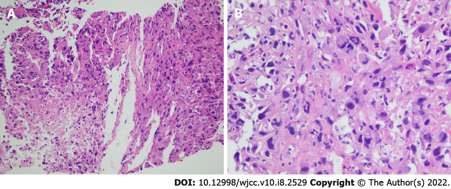
Figure 1 Hematoxylin and eosin staining photomicrographs of a right lung tumor tissue biopsy.
A: Original magnification (×20); B: Original magnification (×40).
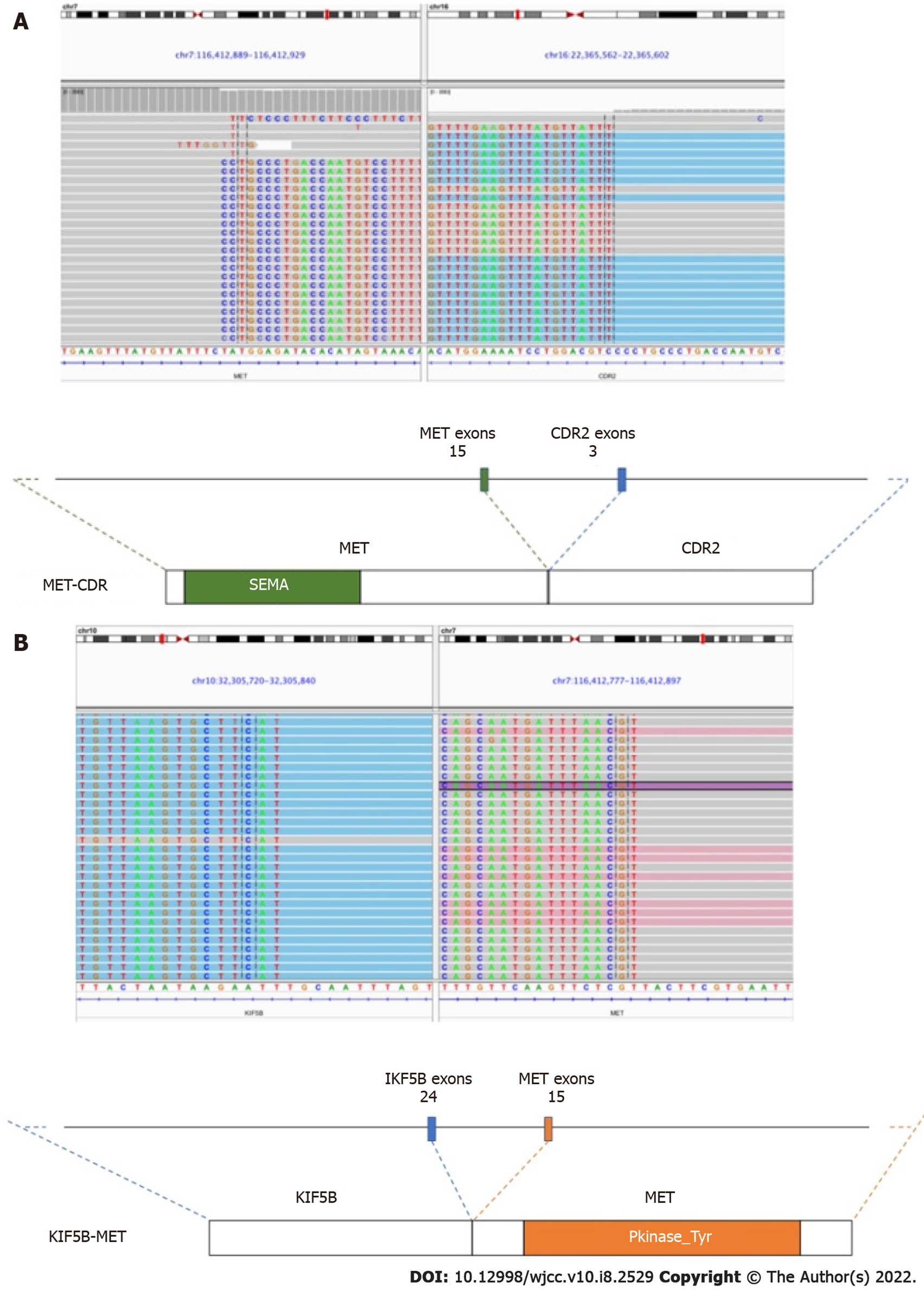
Figure 2 Next-generation sequencing revealed two concurrent MET fusions with different fusion partners, MET-CDR2 and KIF5B-MET.
A: Images from the Integrative Genomics Viewer demonstrating the chromosomal rearrangement involving MET (chromosome 7, sequencing reads with gray background) and CDR2 (chromosome 16, sequencing reads with blue background); B: KIF5B (chromosome 10, sequencing reads with blue background). Illustrations below demonstrate the protein structure resulting from the gene fusions indicating the breakpoints of the nearby exons.
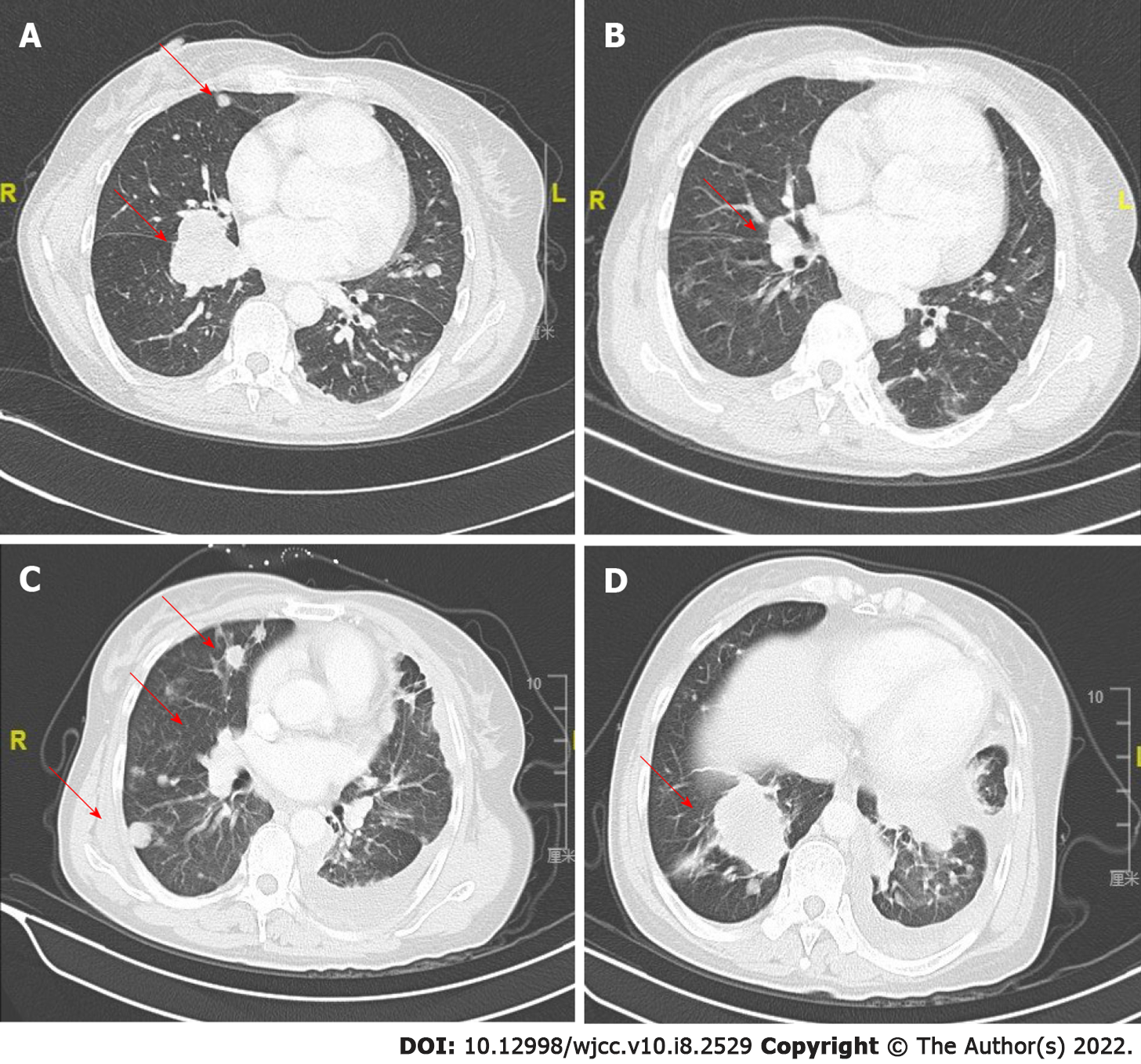
Figure 3 Clinical efficacy of crizotinib treatment in a patient with KIF5B-MET and MET-CDR2-rearranged, poorly differentiated lung cancer.
A: Thoracic computed tomographic image at baseline; B: 1 mo after initiating crizotinib therapy; C and D: After disease progression and another 2 mo later in May, 2019.
- Citation: Liu LF, Deng JY, Lizaso A, Lin J, Sun S. Effective response to crizotinib of concurrent KIF5B-MET and MET-CDR2-rearranged non-small cell lung cancer: A case report. World J Clin Cases 2022; 10(8): 2529-2536
- URL: https://www.wjgnet.com/2307-8960/full/v10/i8/2529.htm
- DOI: https://dx.doi.org/10.12998/wjcc.v10.i8.2529


