Copyright
©The Author(s) 2022.
World J Clin Cases. Mar 16, 2022; 10(8): 2491-2496
Published online Mar 16, 2022. doi: 10.12998/wjcc.v10.i8.2491
Published online Mar 16, 2022. doi: 10.12998/wjcc.v10.i8.2491
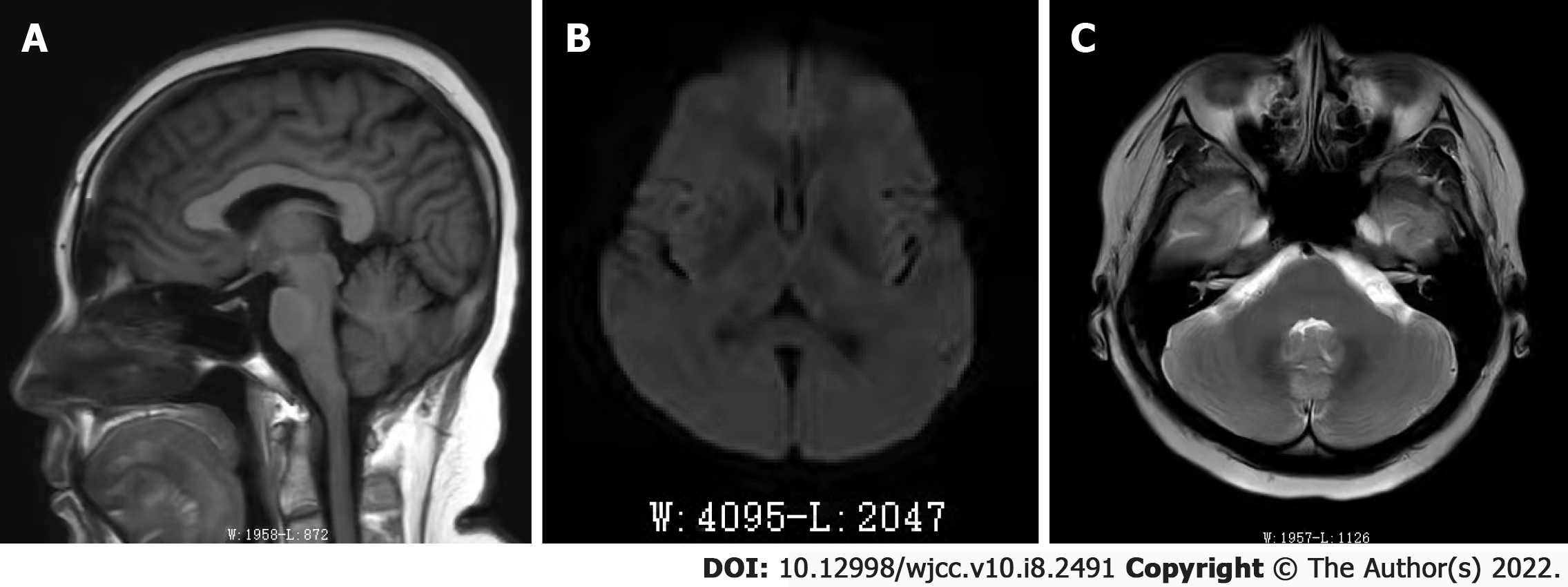
Figure 1 Brain magnetic resonance images of the patient.
A: Axial view of T1-weighted image shows no brain dysplasia, encephalomalacia or abnormal white matter signal; B: Diffusion-weighted image shows no abnormal signals; C: T2-weighted scan shows that the bilateral internal auditory canal, cochlear, auditory and cranial nerve have no abnormal signals.
- Citation: Liu YZ, Jiang H, Zhao YH, Zhang Q, Hao SC, Bao LP, Wu W, Jia ZB, Jiang HC. Severe tinnitus and migraine headache in a 37-year-old woman treated with trastuzumab for breast cancer: A case report. World J Clin Cases 2022; 10(8): 2491-2496
- URL: https://www.wjgnet.com/2307-8960/full/v10/i8/2491.htm
- DOI: https://dx.doi.org/10.12998/wjcc.v10.i8.2491


