Copyright
©The Author(s) 2022.
World J Clin Cases. Mar 6, 2022; 10(7): 2315-2321
Published online Mar 6, 2022. doi: 10.12998/wjcc.v10.i7.2315
Published online Mar 6, 2022. doi: 10.12998/wjcc.v10.i7.2315
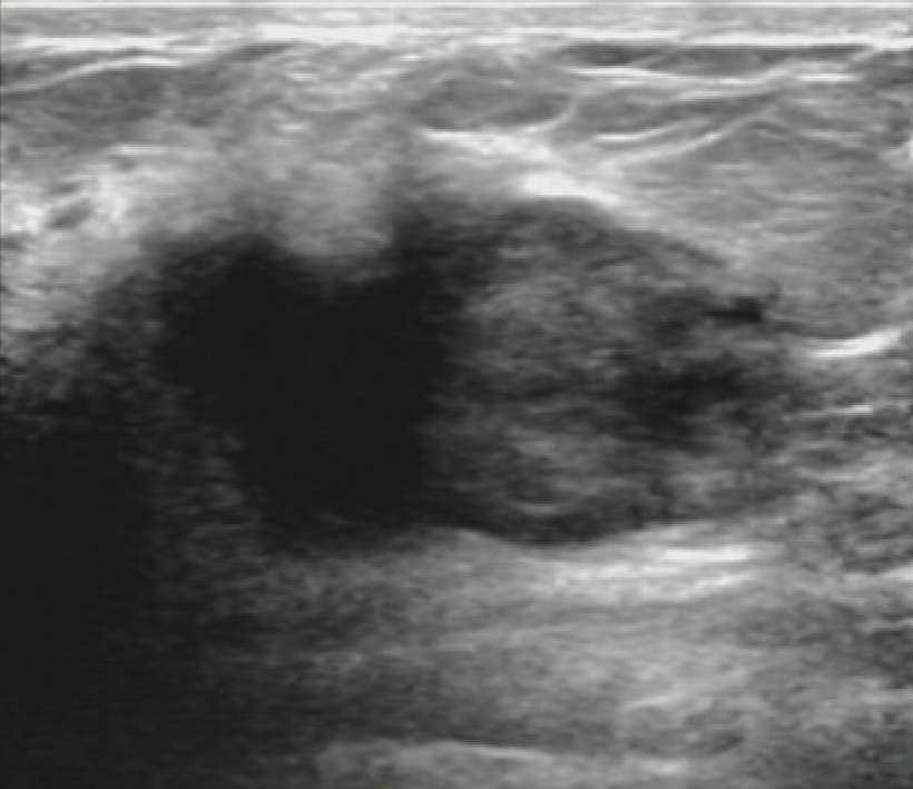
Figure 1 Sonographic image of the patient’s right breast.
A hypoechoic mass, measuring 3 cm 4 cm, was found in the upper inner quadrant.
Figure 2 Positron-emission tomography/magnetic resonance imaging showed contrast enhancement of multiple masses in the upper inner quadrant of the right breast.
A: Magnetic resonance (MR) showed the mass was hyperactive on T1-weighted images; B: MR showed a soft tissue shadow in the right breast on T2-weighted images; C: Positron-emission tomography showed the fluorodeoxyglucose uptake of the right breast enhanced.
Figure 3 Positron-emission tomography/magnetic resonance imaging showed densely enhancing homogeneous masses in the spinal canal at the C7/T1 levels.
A: Magnetic resonance (MR) showed the right side of C7/T1 vertebral body and its accessories as a low signal on T1-weighted images; B: MR showed soft tissue shadow at the C7/T1 levels on T2-weighted images; C: Positron-emission tomography showed the fluorodeoxyglucose uptake enhanced at the C7/T1 levels.
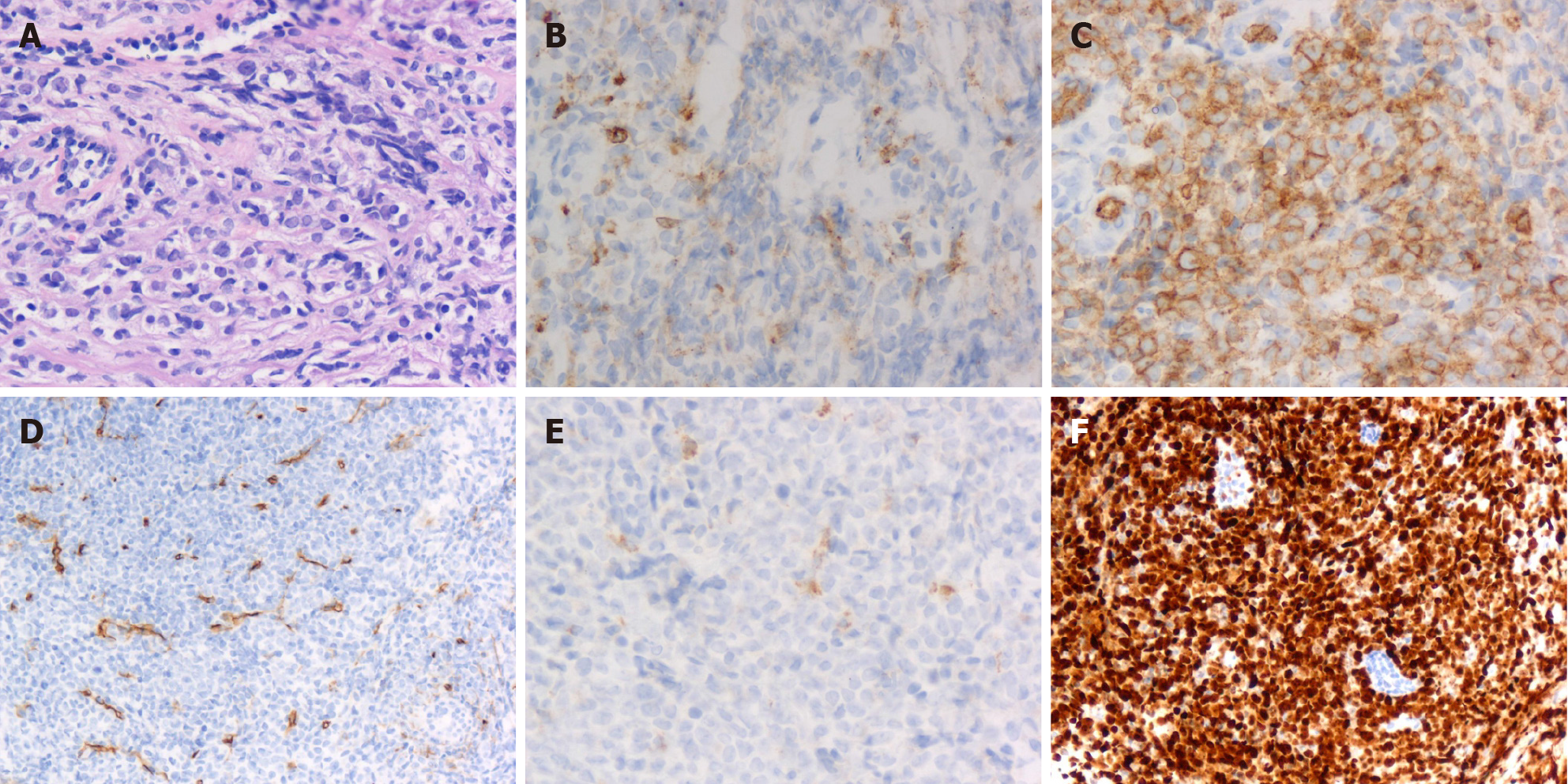
Figure 4 Granulocytic sarcoma of the breast in pathological study.
A: Histology showed a diffuse infiltrate of medium-sized cells with finely dispersed chromatin and small distinct nucleoli with scant cytoplasm (hematoxylin and eosin, × 200 magnification); B-F: Granulocytic sarcoma of the breast was positive for myeloperoxidase (B: × 200 magnification), CD117 (C: × 200 magnification), CD34 (D: × 200 magnification), CD68 (E: × 200 magnification) and Ki67 proliferation index approximately 80% (F: × 200 magnification).
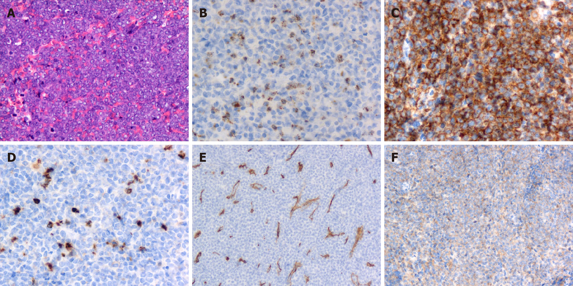
Figure 5 Granulocytic sarcoma of the dorsal spine in pathological study.
A: Histology showed a diffuse infiltrate of intermediate-sized cells with scant cytoplasm and small nucleoli (hematoxylin and eosin, × 200 magnification); B-F: Granulocytic sarcoma of the dorsal spine was positive for myeloperoxidase (B: × 200 magnification), CD117 (C: × 200 magnification), lysozyme (D: × 200 magnification), CD34 (E: × 200 magnification) and CD38 (F: × 200 magnification).
- Citation: Li Y, Xie YD, He SJ, Hu JM, Li ZS, Qu SH. Breast and dorsal spine relapse of granulocytic sarcoma after allogeneic stem cell transplantation for acute myelomonocytic leukemia: A case report. World J Clin Cases 2022; 10(7): 2315-2321
- URL: https://www.wjgnet.com/2307-8960/full/v10/i7/2315.htm
- DOI: https://dx.doi.org/10.12998/wjcc.v10.i7.2315


