Copyright
©The Author(s) 2022.
World J Clin Cases. Mar 6, 2022; 10(7): 2222-2228
Published online Mar 6, 2022. doi: 10.12998/wjcc.v10.i7.2222
Published online Mar 6, 2022. doi: 10.12998/wjcc.v10.i7.2222
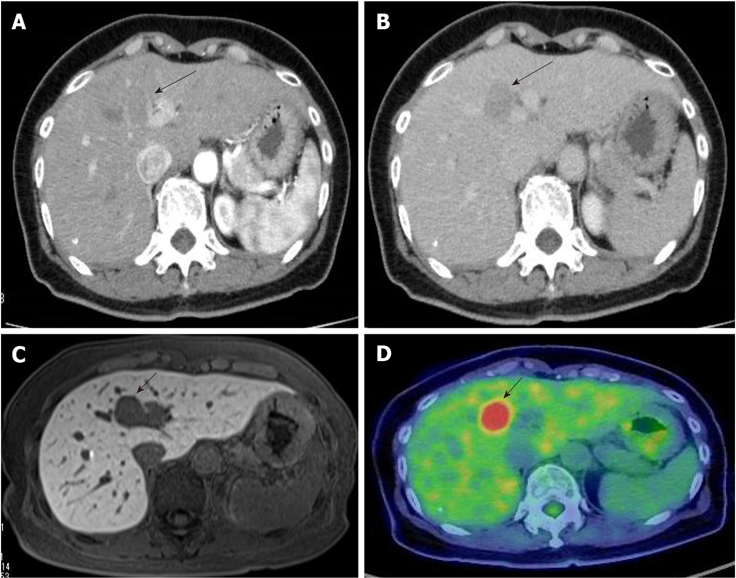
Figure 1 Abdominal computed tomography.
A: Peripheral contrast enhancement was observed in the atrial phase (arrow); B: In the delayed phase, the contrast of the mass appeared lower than the surrounding liver tissue (arrow); C: The decreased uptake of Gadoxetate sodium was found (arrow); D: Positron emission tomography/computed tomography showed significant accumulation at the mass (arrow).
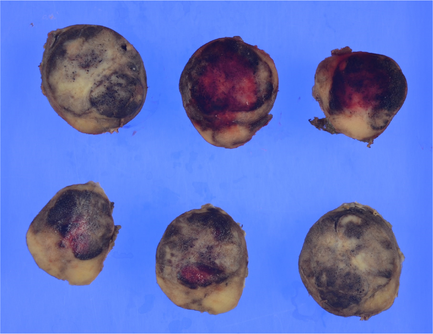
Figure 2 Macroscopic findings of the excised specimen.
The specimen was a 2.5 cm × 2.0 cm × 3.0 cm yellowish-white mass with hemorrhage.
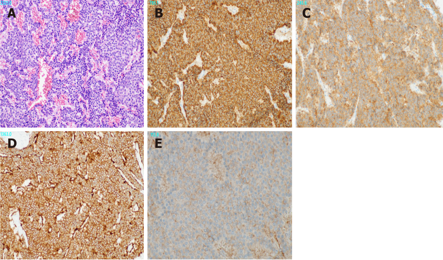
Figure 3 Microscopic findings of the tumor.
A: Atypical cells with small round nuclei and eosinophilic cytoplasm were arranged in an alveolar, reticular, or trabecular pattern. (Hematoxylin and eosin staining results; × 200); B: The tumor cells were positive for Chromogranin A (× 200); C: They were positive for Synaptophysin (× 200); D: They were positive for somatostatin receptor (SSTR)2 (× 200); E: They were positive for SSTR5 (× 400).
- Citation: Akabane M, Kobayashi Y, Kinowaki K, Okubo S, Shindoh J, Hashimoto M. Primary hepatic neuroendocrine neoplasm diagnosed by somatostatin receptor scintigraphy: A case report. World J Clin Cases 2022; 10(7): 2222-2228
- URL: https://www.wjgnet.com/2307-8960/full/v10/i7/2222.htm
- DOI: https://dx.doi.org/10.12998/wjcc.v10.i7.2222


