Copyright
©The Author(s) 2022.
World J Clin Cases. Feb 26, 2022; 10(6): 2007-2014
Published online Feb 26, 2022. doi: 10.12998/wjcc.v10.i6.2007
Published online Feb 26, 2022. doi: 10.12998/wjcc.v10.i6.2007
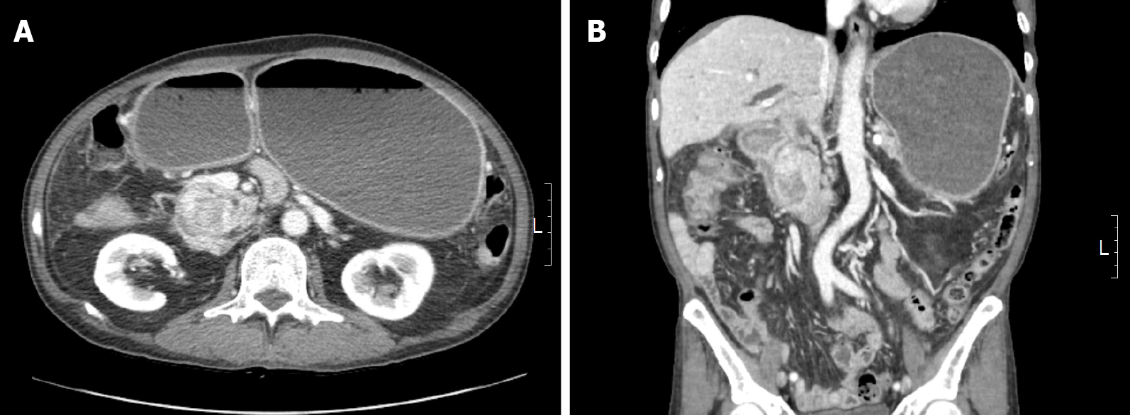
Figure 1 Radiologic findings.
A, B: Abdominal computed tomography scan demonstrated a heterogeneously enhanced mass in the pancreaticoduodenal groove with duodenal obstruction.
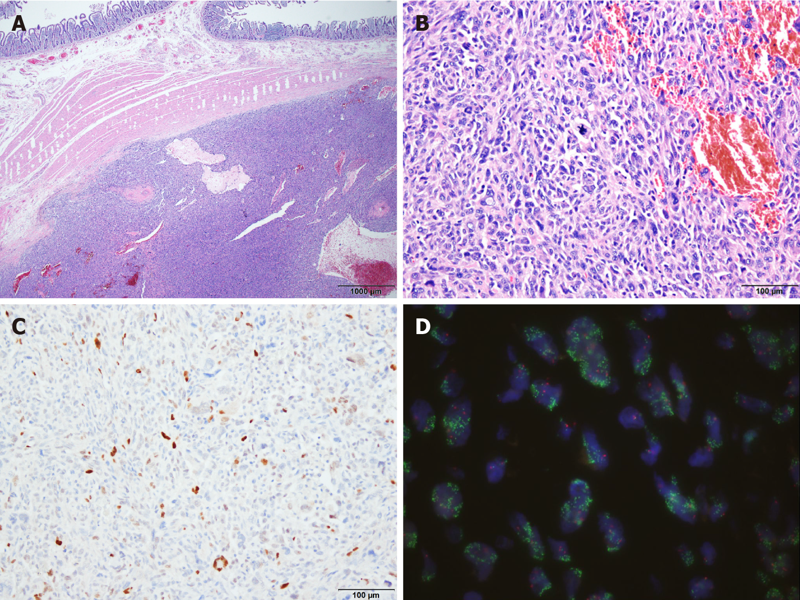
Figure 2 Features of tumor cells.
A: The tumor was located in the submucosal layer of the duodenum (Hematoxylin-and-eosin stain, ×20); B: At a higher magnification, undifferentiated tumor cells were shown to have marked nuclear atypia with brisk mitotic activity (Hematoxylin-and-eosin stain, ×200); C: Immunohistochemistry revealed positivity for MDM2 in the tumor cells (Immunohistochemistry, ×200); D: MDM2 amplification was detected by MDM2/CEN12 fluorescence in situ hybridization assay (MDM2-green signals, CEN12-red signals, ×1000).
- Citation: Kim NI, Lee JS, Choi C, Nam JH, Choi YD, Kim HJ, Kim SS. Primary duodenal dedifferentiated liposarcoma: A case report and literature review. World J Clin Cases 2022; 10(6): 2007-2014
- URL: https://www.wjgnet.com/2307-8960/full/v10/i6/2007.htm
- DOI: https://dx.doi.org/10.12998/wjcc.v10.i6.2007


