Copyright
©The Author(s) 2022.
World J Clin Cases. Feb 26, 2022; 10(6): 1952-1960
Published online Feb 26, 2022. doi: 10.12998/wjcc.v10.i6.1952
Published online Feb 26, 2022. doi: 10.12998/wjcc.v10.i6.1952
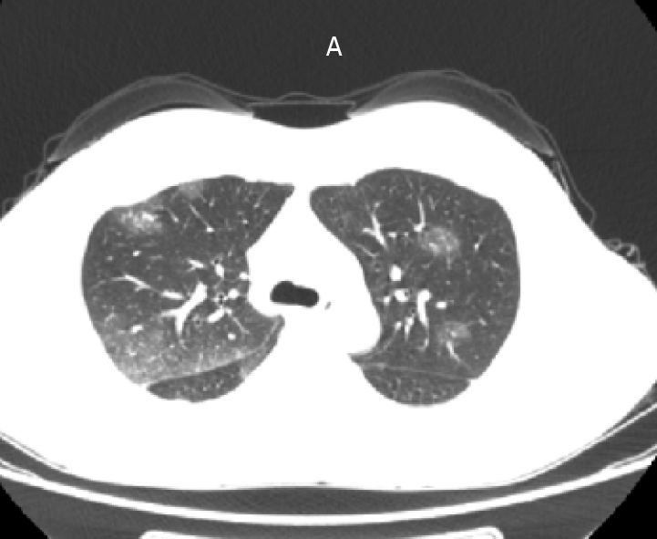
Figure 1
Computed tomography of the chest and upper abdomen reveals bilateral pleural effusion, with suspected involvement of inflammatory exudates.
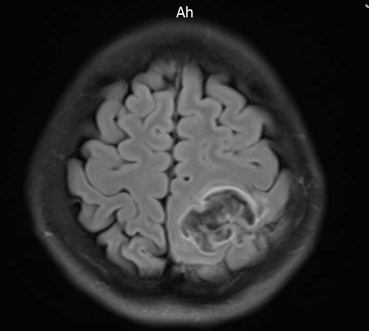
Figure 2
Abnormal signals on the left side at the fronto-parietal junction, indicating the formation of a hematoma.
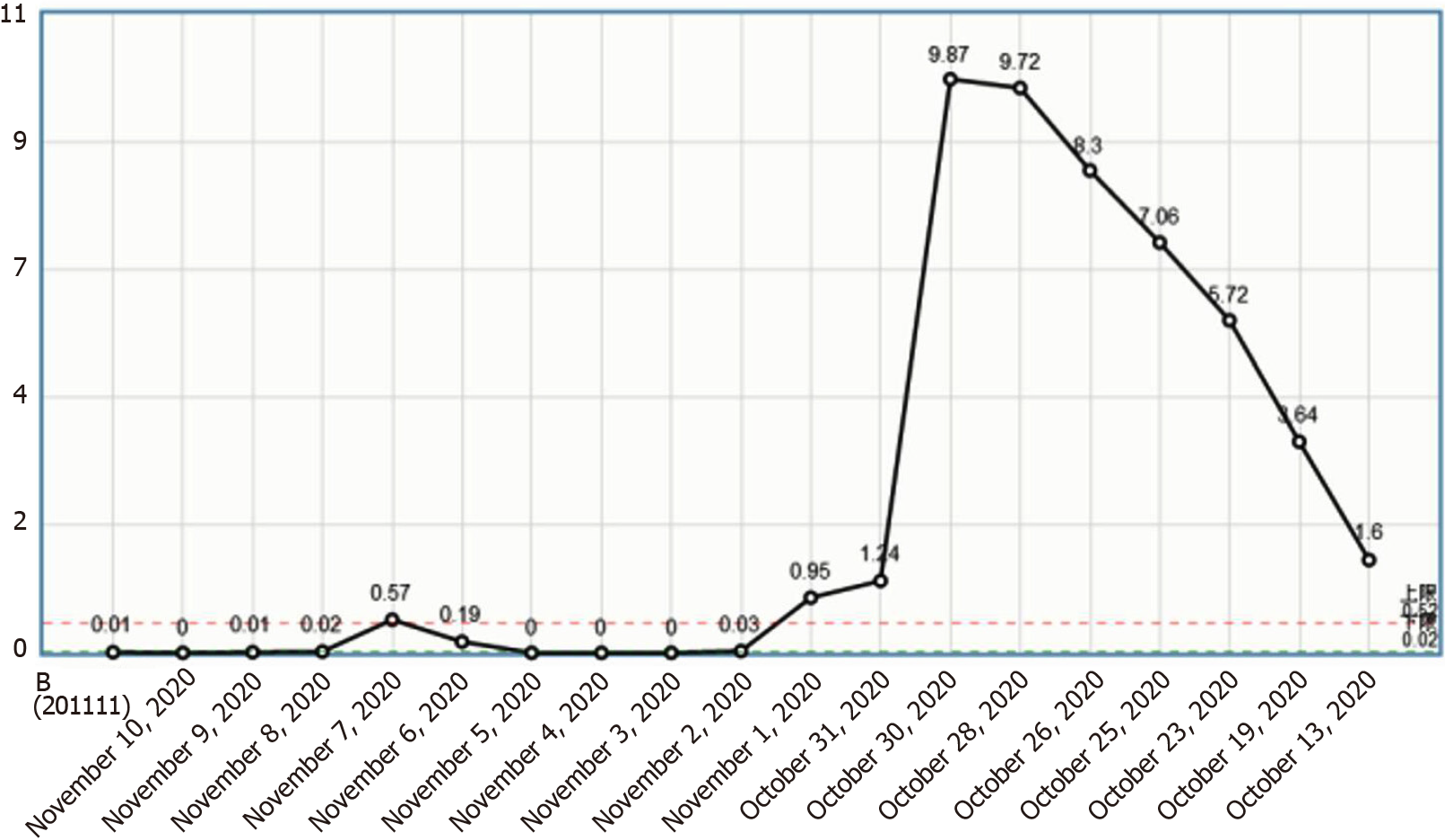
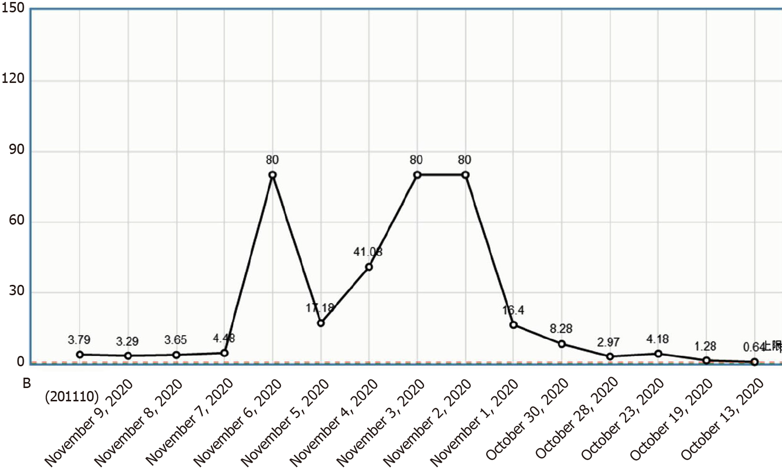
Figure 3
Change curve of the eosinophil count.
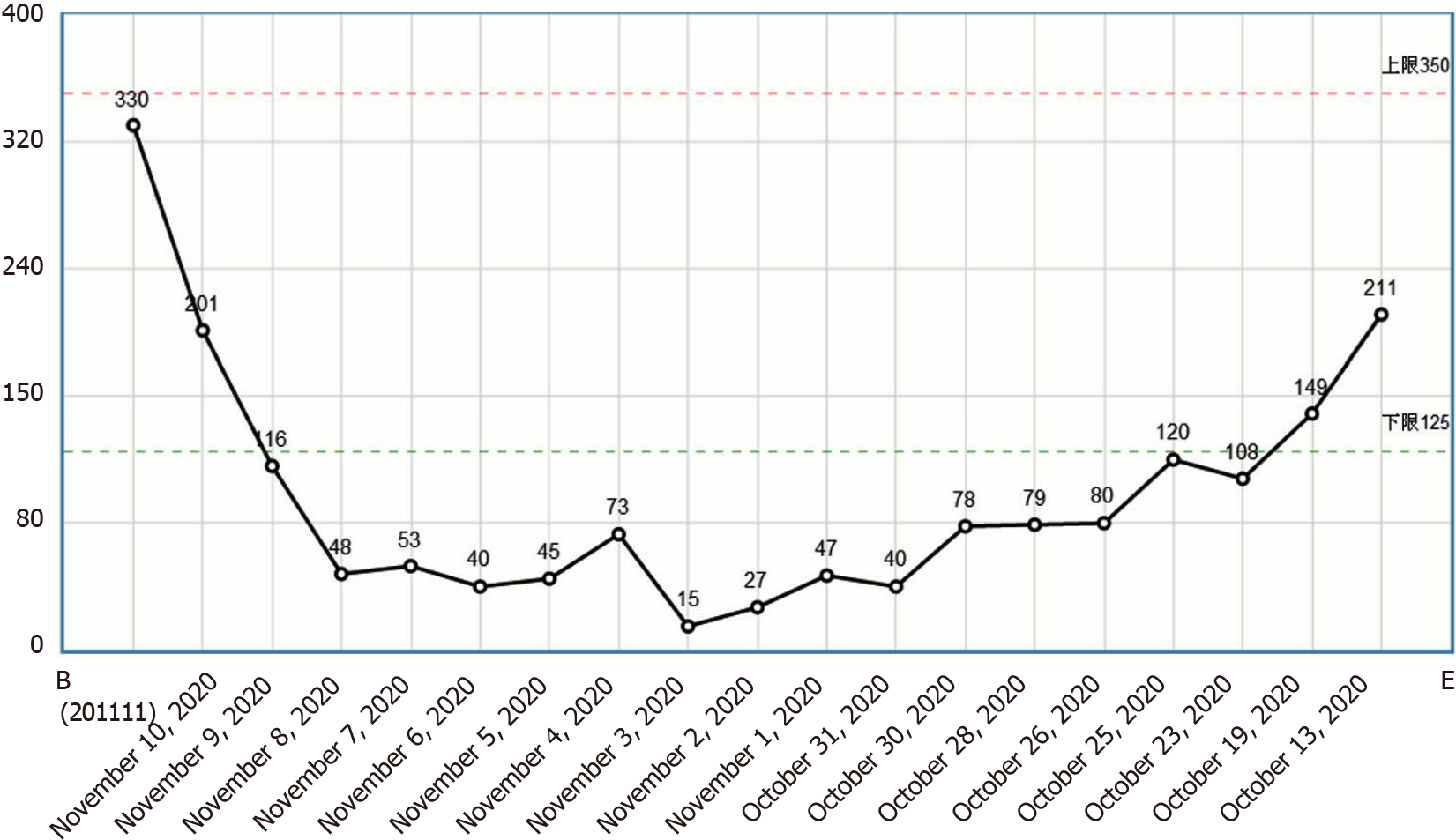
Figure 4
Change curve of the platelet count.
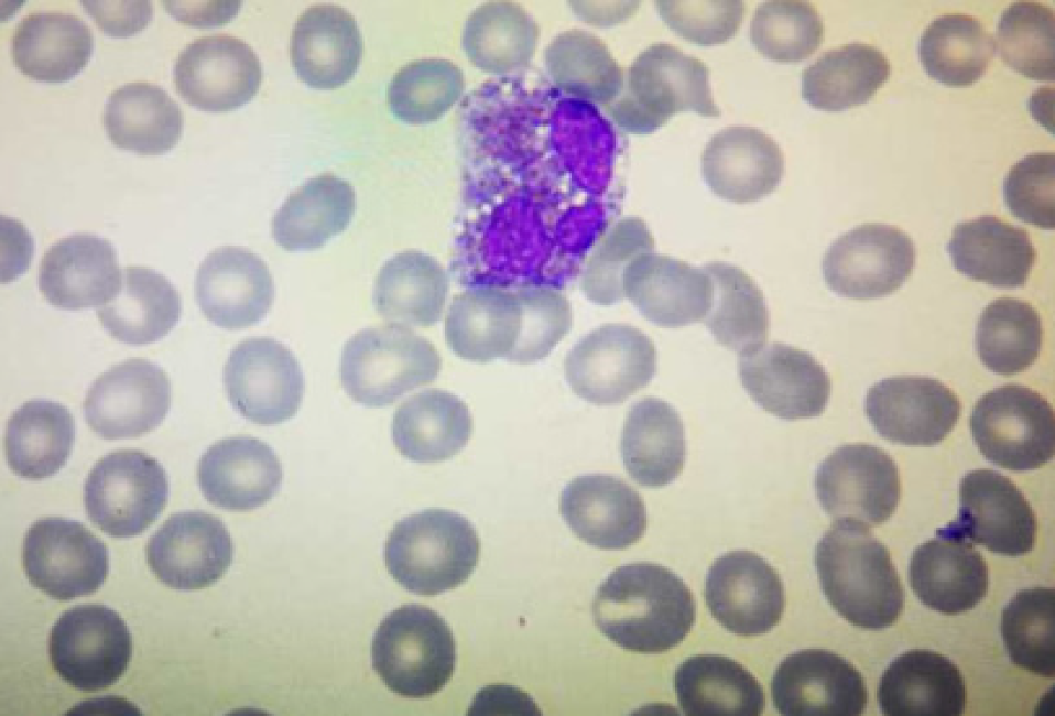
Figure 5
Eosinophil degranulation.
Figure 6 D-dimer change curve.
- Citation: Su WQ, Fu YZ, Liu SY, Cao MJ, Xue YB, Suo FF, Liu WC. Eosinophilia complicated with venous thromboembolism: A case report. World J Clin Cases 2022; 10(6): 1952-1960
- URL: https://www.wjgnet.com/2307-8960/full/v10/i6/1952.htm
- DOI: https://dx.doi.org/10.12998/wjcc.v10.i6.1952


