Copyright
©The Author(s) 2022.
World J Clin Cases. Feb 16, 2022; 10(5): 1689-1696
Published online Feb 16, 2022. doi: 10.12998/wjcc.v10.i5.1689
Published online Feb 16, 2022. doi: 10.12998/wjcc.v10.i5.1689
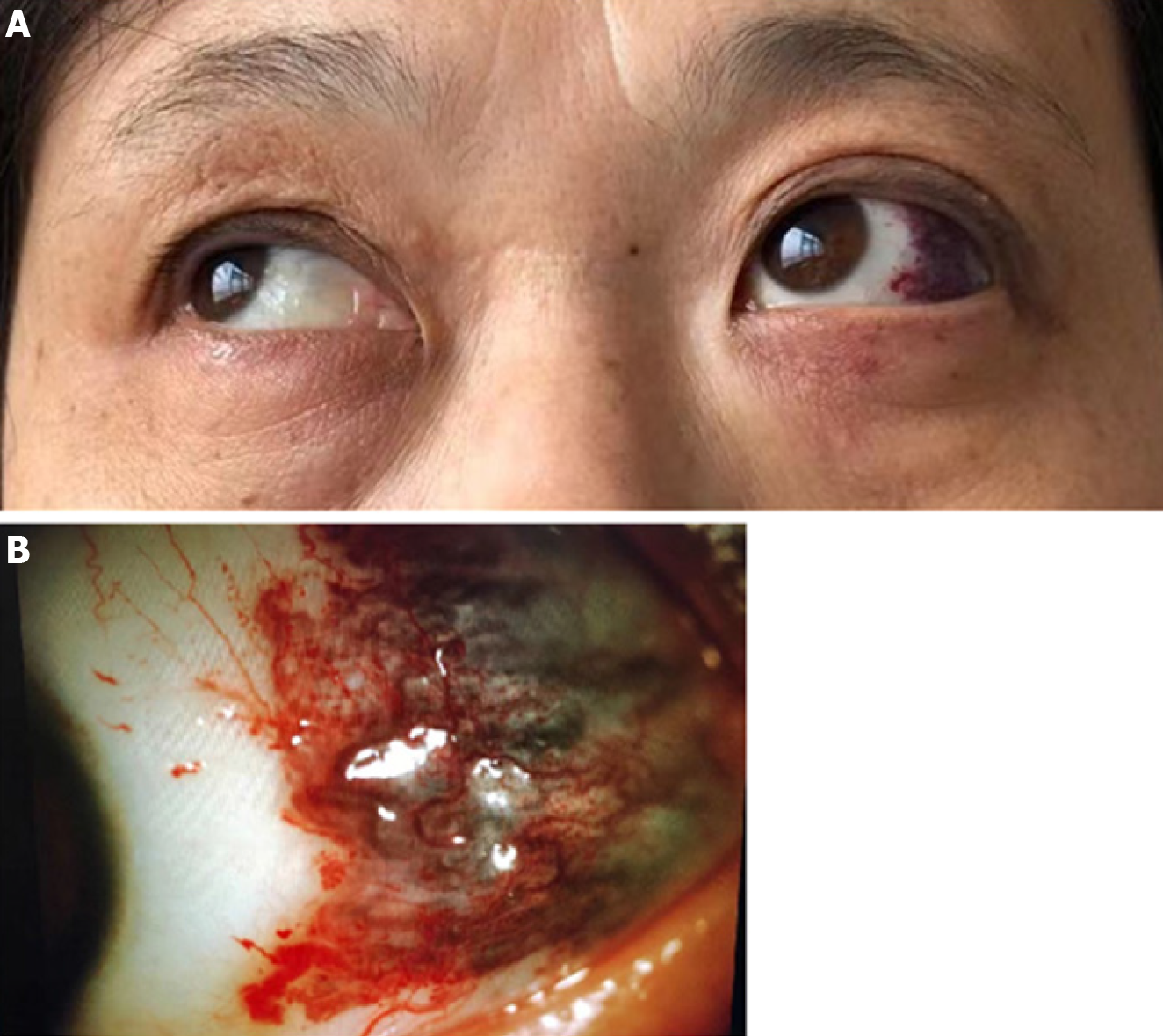
Figure 1 The slit-lamp examination of left eye.
A: The left eyeball protruded forward; B: The slit-lamp examination found that her left conjunctiva was not congestible and curled blood vessels were seen under the conjunctiva in the temporal side, which color was dark purple, with a range of about 1 cm × 1 cm.
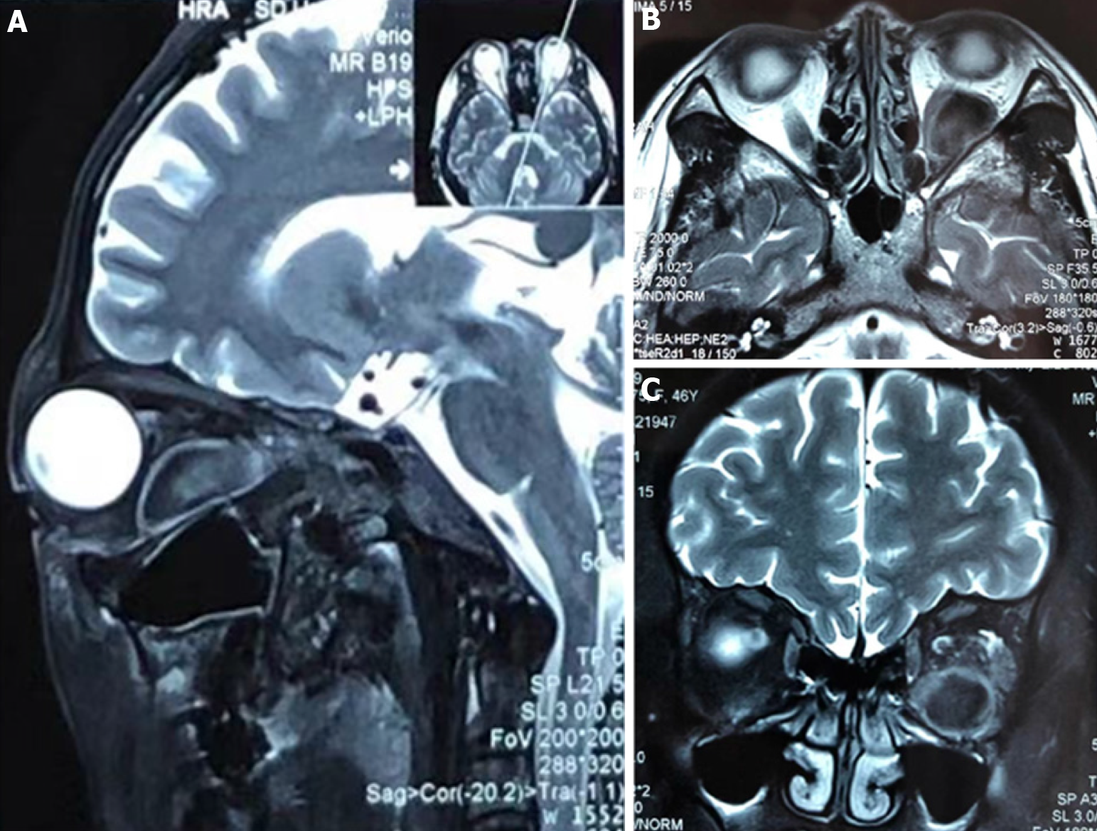
Figure 2 Magnetic resonance imaging scans of orbits.
A-C: Magnetic resonance imaging scans of orbits showed that the left eye was protruding, and an elliptical long-short T1 long-short T2 signal focus was observed in the lateral optic nerve of the left orbital muscle cone, with smooth edges and low signal on DWI. The left hyperdense retrobulbar mass displaced optic nerve superomedial.
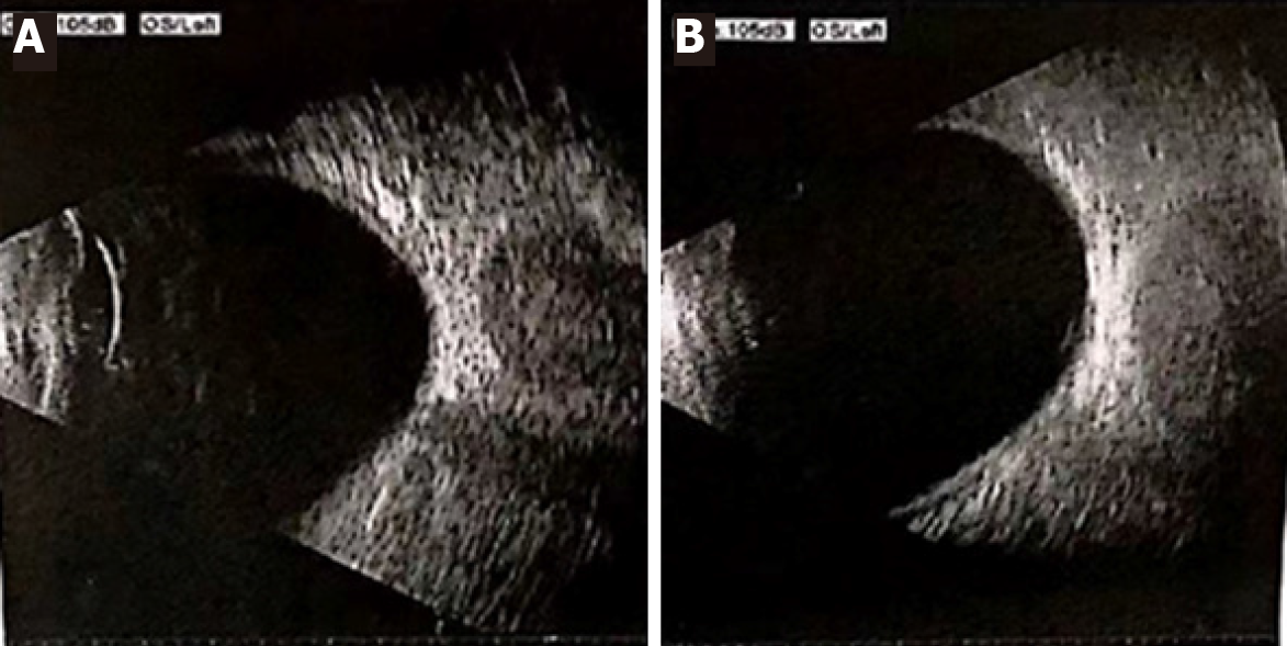
Figure 3 Ocular ultrasound of the left eye.
A, B: Ocular Ultrasound showed that the echo of the posterior eyeball mass of the left eye is uneven, and it disappears while the gain reduced, suggesting there was a goitre of the left orbit.
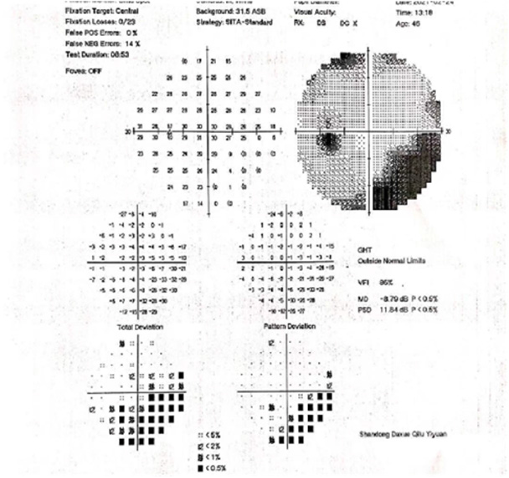
Figure 4 Visual field examination.
Visual field examination revealed a visual field defect below the temporal of the left eye.
- Citation: Lei JY, Wang H. Bulbar conjunctival vascular lesion combined with spontaneous retrobulbar hematoma: A case report. World J Clin Cases 2022; 10(5): 1689-1696
- URL: https://www.wjgnet.com/2307-8960/full/v10/i5/1689.htm
- DOI: https://dx.doi.org/10.12998/wjcc.v10.i5.1689


