Copyright
©The Author(s) 2022.
World J Clin Cases. Feb 16, 2022; 10(5): 1684-1688
Published online Feb 16, 2022. doi: 10.12998/wjcc.v10.i5.1684
Published online Feb 16, 2022. doi: 10.12998/wjcc.v10.i5.1684
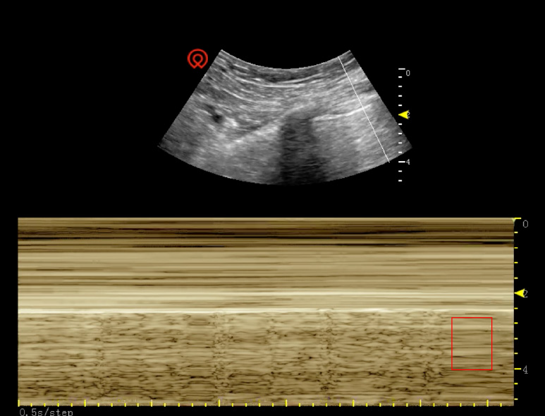
Figure 1 Ultrasound showed an advection level sign on M-mode during the operation.
Red square indicates the advection level sign on M-mode, whereas no B line could be detected on ultrasonography.
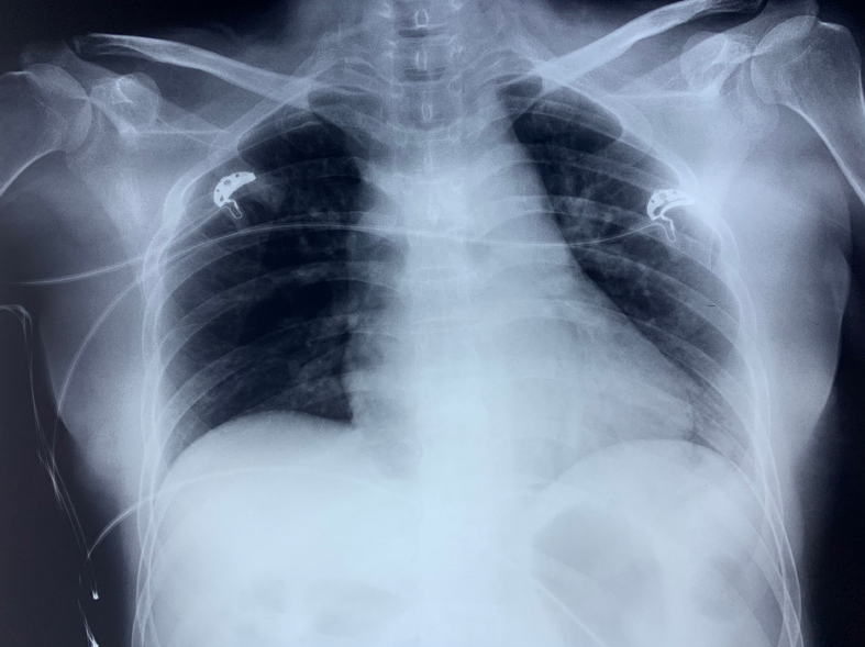
Figure 2 Bedside chest X-ray result after the patient had been transferred to the urosurgery ward.
No evidence of pneumothorax or lib injury was found after the lung dilation.
- Citation: Zhao Y, Xue XQ, Xia D, Xu WF, Liu GH, Xie Y, Ji ZG. Pneumothorax during retroperitoneal laparoscopic partial nephrectomy in a lupus nephritis patient: A case report. World J Clin Cases 2022; 10(5): 1684-1688
- URL: https://www.wjgnet.com/2307-8960/full/v10/i5/1684.htm
- DOI: https://dx.doi.org/10.12998/wjcc.v10.i5.1684


