Copyright
©The Author(s) 2022.
World J Clin Cases. Feb 16, 2022; 10(5): 1675-1683
Published online Feb 16, 2022. doi: 10.12998/wjcc.v10.i5.1675
Published online Feb 16, 2022. doi: 10.12998/wjcc.v10.i5.1675
Figure 1 Abdominal ultrasonography of the mass.
A giant hyperechoic mass filling the abdomen was presented on grey-scale ultrasound. The mass had a relative clear margin and internal septas.
Figure 2 Abdominal computed tomography in the axial plane.
Computed tomography imaging showed a giant homogenous mass, mainly consisting of fatty tissue measuring 16.6 cm × 28.6 cm with thin septa, pushing the peritoneal containing such as bowel loops and uterus to the right part of abdomen.
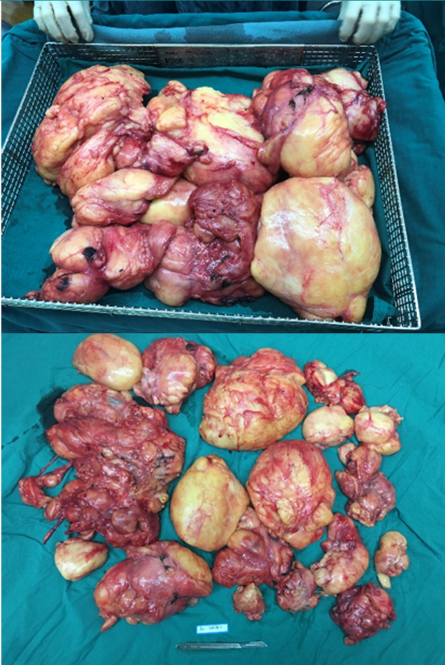
Figure 3 Macroscopic view of extracted retroperitoneal lipoma.
During the operation, a bulky yellowish tumor, originating from the left retroperitoneal region, was found to occupy the retroperitoneum. The mass weighted 7.126 kg.
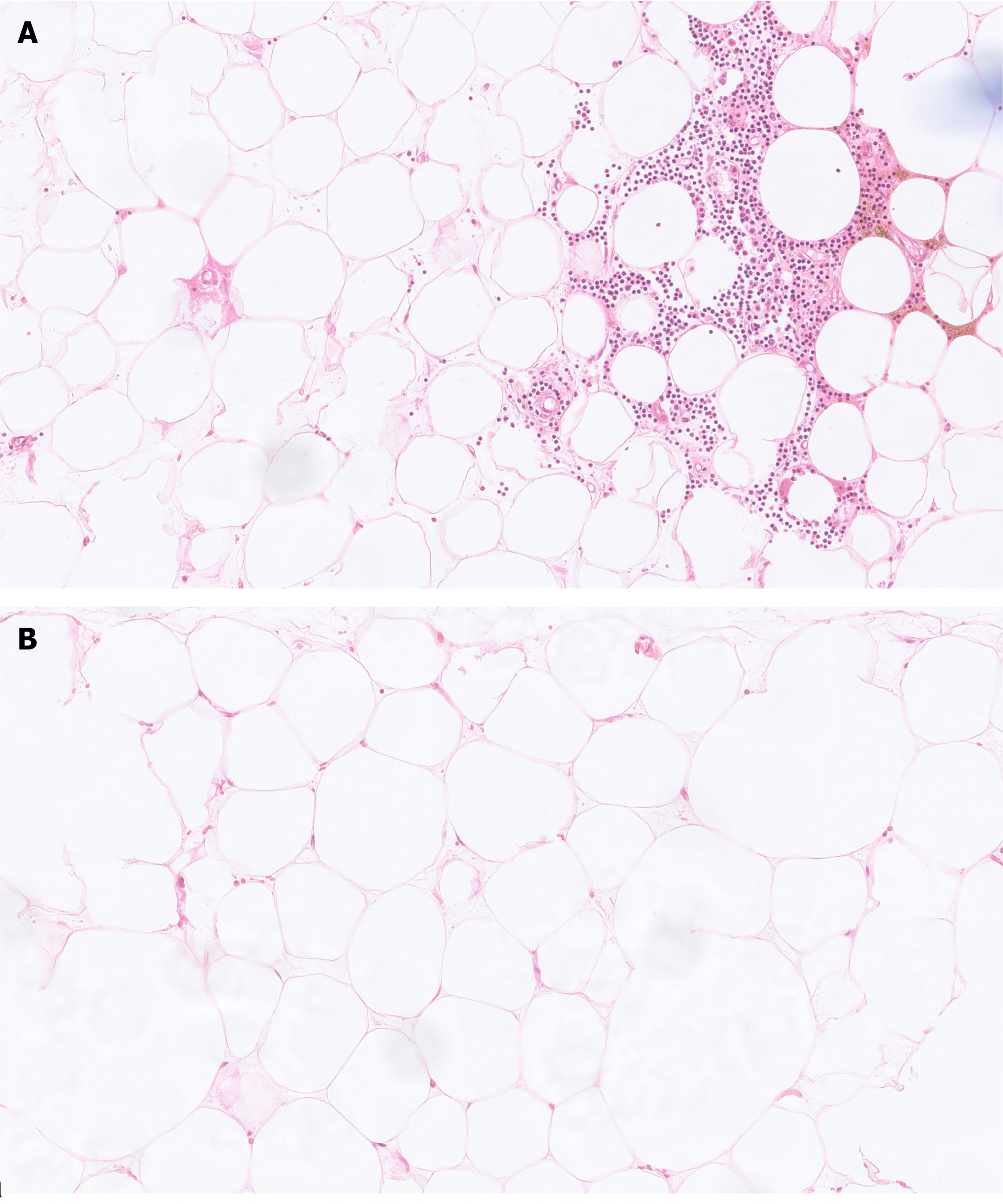
Figure 4 Microscopical picture of the extracted tumors (H&E, 20 ×).
A: Myelolipoma was composed of mature adipose adipocytes and hematopoietic cells, without necrosis, atypia, and hyperchromatic cells; B: Conventional lipoma was composed of mature adipocytes.
- Citation: Chen ZY, Chen XL, Yu Q, Fan QB. Giant retroperitoneal lipoma presenting with abdominal distention: A case report and review of the literature. World J Clin Cases 2022; 10(5): 1675-1683
- URL: https://www.wjgnet.com/2307-8960/full/v10/i5/1675.htm
- DOI: https://dx.doi.org/10.12998/wjcc.v10.i5.1675


