Copyright
©The Author(s) 2022.
World J Clin Cases. Feb 16, 2022; 10(5): 1654-1666
Published online Feb 16, 2022. doi: 10.12998/wjcc.v10.i5.1654
Published online Feb 16, 2022. doi: 10.12998/wjcc.v10.i5.1654
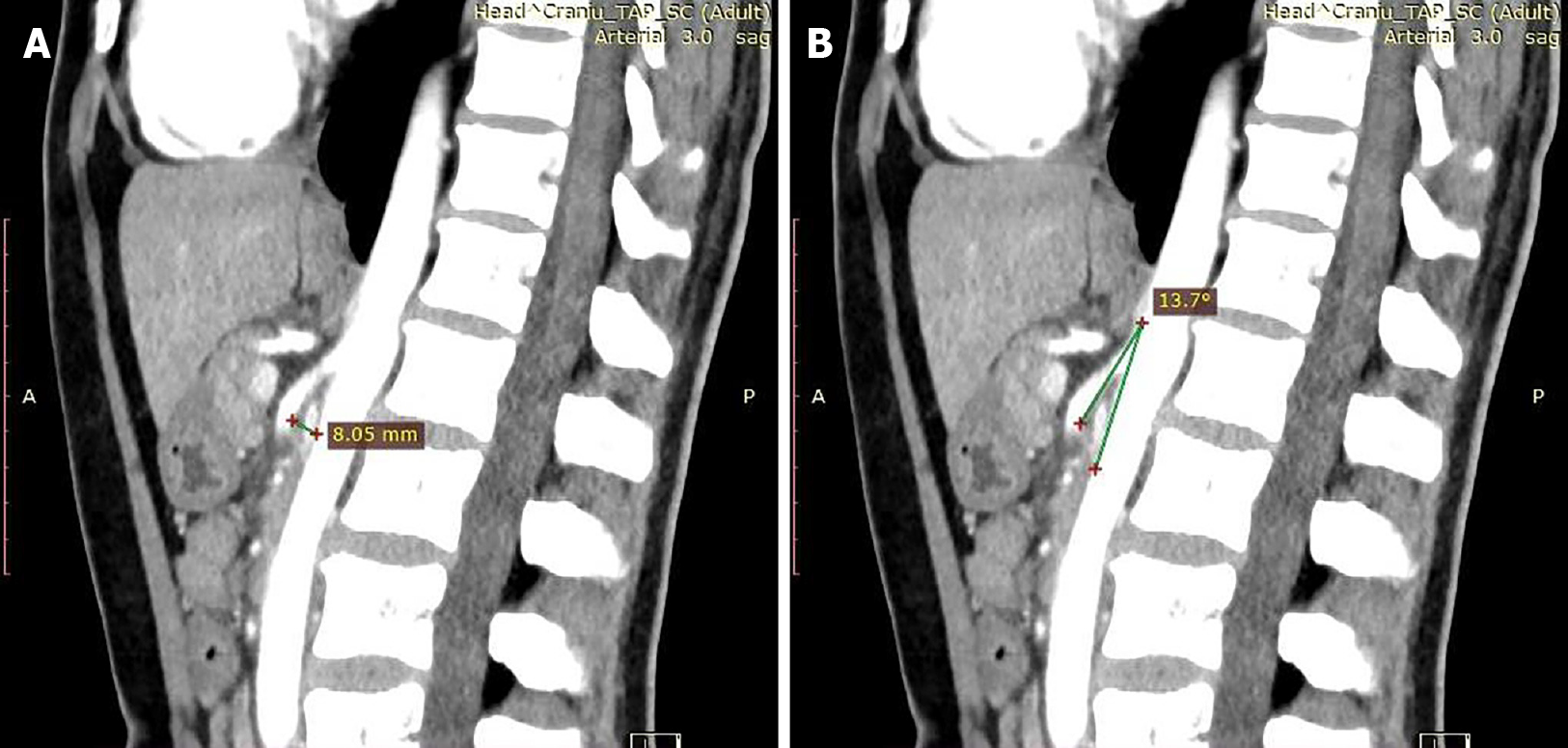
Figure 1 Angio-computed tomography images.
A: Reduced distance between the superior mesenteric artery (SMA) and the anterior wall of the aorta up to a maximum of 8.05 mm; B: Emergence at a sharp angle of 13.7° of the SMA.
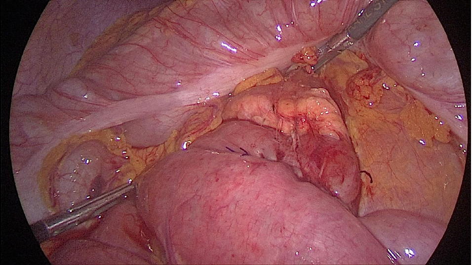
Figure 2 Exploratory laparoscopy.
A: Dilated duodenum in the first and second parts; B: A collapsed third part beyond the mesenteric pedicle.
Figure 3 Laparoscopic duodenojejunostomy.
A: Threaded spotting of the two enteral segments for the anastomosis; B: Side-to-side duodenojejunostomy using a 60 mm endoscopic liner articulated stapler.
Figure 4 Final aspect of the anastomosis after closing the common enterotomy.
Figure 5 Postoperative aspect of the abdomen at 1 mo evaluation, with 10 kg weight gain.
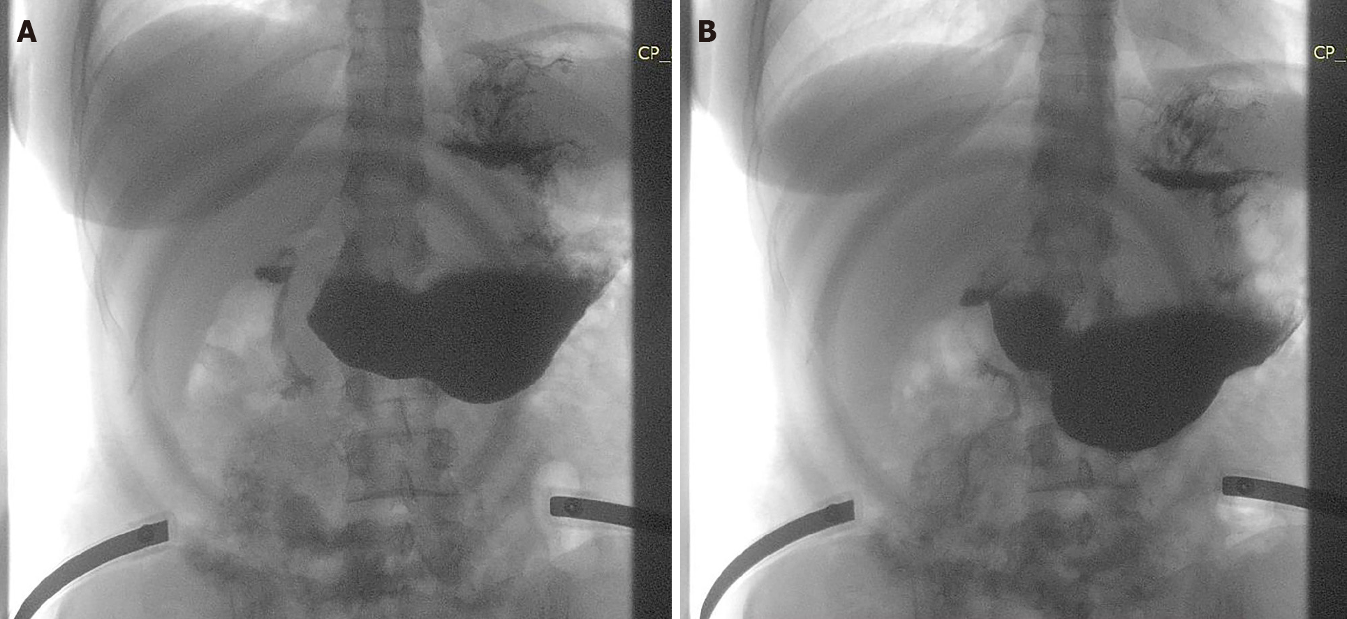
Figure 6 Oral gastrografin study.
A: Free passage of the contrast through the anastomosis in the small bowel; B: Contraction of the stomach and duodenum.
- Citation: Apostu RC, Chira L, Colcear D, Lebovici A, Nagy G, Scurtu RR, Drasovean R. Wilkie’s syndrome as a cause of anxiety-depressive disorder: A case report and review of literature. World J Clin Cases 2022; 10(5): 1654-1666
- URL: https://www.wjgnet.com/2307-8960/full/v10/i5/1654.htm
- DOI: https://dx.doi.org/10.12998/wjcc.v10.i5.1654


