Copyright
©The Author(s) 2022.
World J Clin Cases. Feb 16, 2022; 10(5): 1645-1653
Published online Feb 16, 2022. doi: 10.12998/wjcc.v10.i5.1645
Published online Feb 16, 2022. doi: 10.12998/wjcc.v10.i5.1645
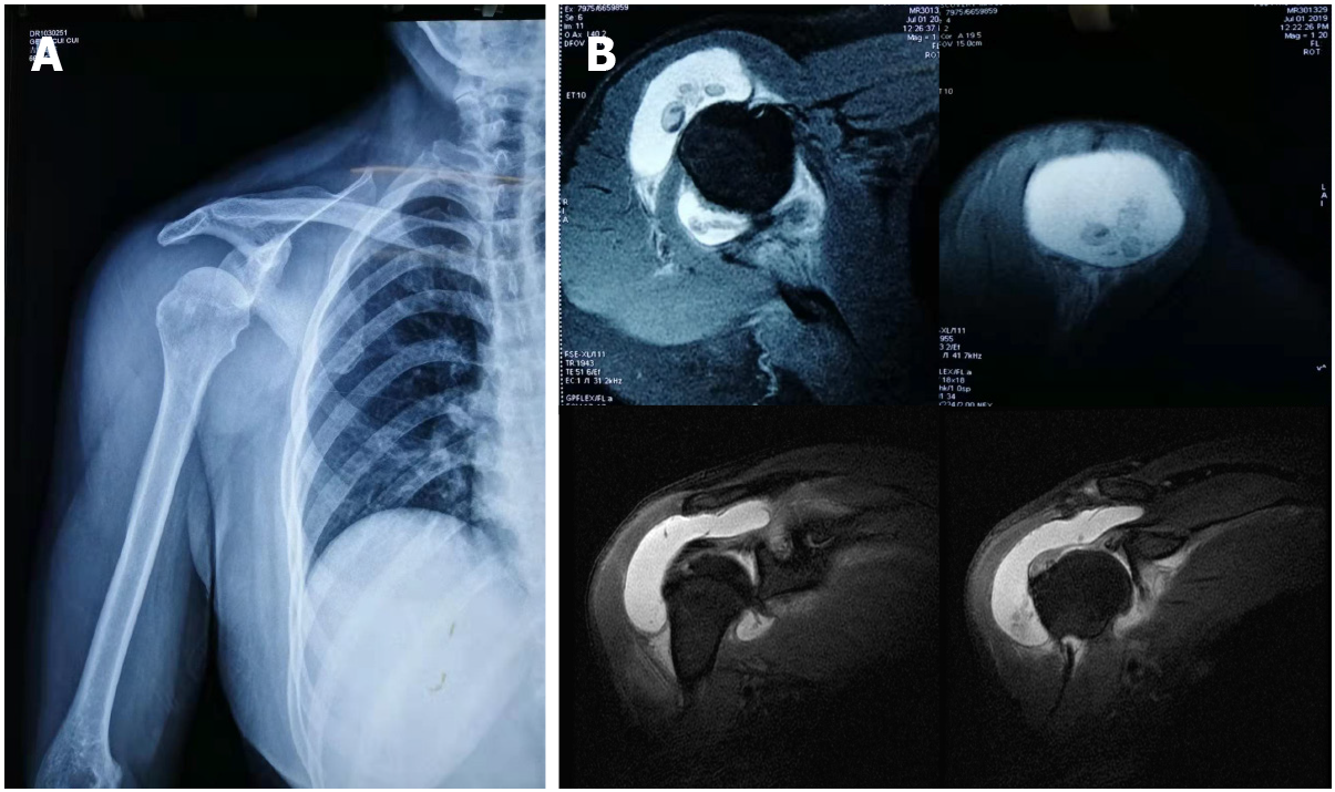
Figure 1 Imaging examinations of case one.
A: Plain radiograph in anteroposterior view of the right shoulder, showing subluxation of the humeral head; B: Magnetic resonance imaging examination of the shoulder joint, showing multiple intra-articular loose bodies and joint effusion.
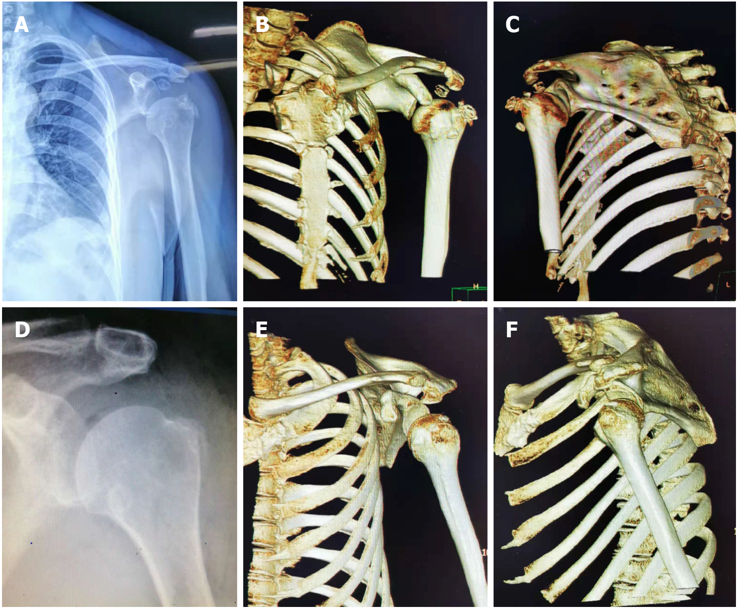
Figure 2 Imaging examinations of case two.
A–C: Plain radiographs and three-dimensional computed tomography (3D-CT) reconstruction of the left shoulder, showing multiple intra-articular loose bodies and the subluxation of the humeral head; D–F: Postoperative radiographic re-examination and 3D-CT reconstruction showed no loose bodies in the subacromial space. The humeral head returned to a normal anatomical relationship.
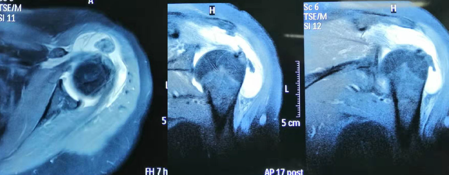
Figure 3
Magnetic resonance imaging of the shoulder joint showed multiple intra-articular loose bodies and joint effusion.
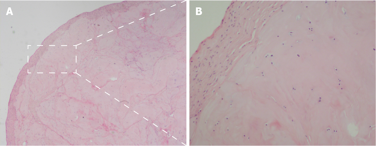
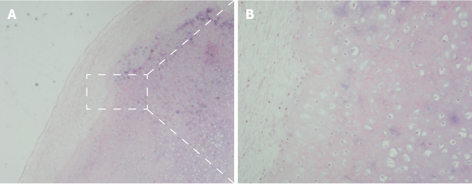
Figure 4 Pathological examination of case one.
A: Lobulated areas of hyaline cartilage just below the synovial surface were easily identified by hematoxylin-eosin staining (magnification: 20 ×); B: Chondrocytes were found clustered together and were not uniformly distributed throughout the ground substance (magnification: 100 ×).
Figure 5 Pathological examination of case two.
A: Fragments of articular cartilage or subchondral lamellar bone were present in loose bodies identified by hematoxylin-eosin staining (magnification: 20 ×); B: Chondrocytes were found in the zonal and ring-like units together and were uniformly distributed throughout the ground substance (magnification: 100 ×).
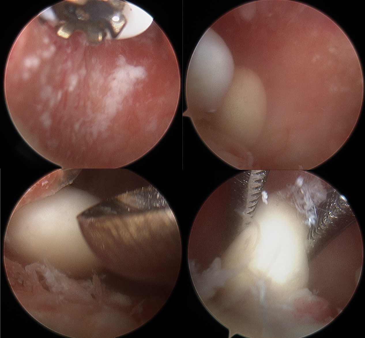
Figure 6
From the standard posterior portal, the arthroscopic view revealed a large number of cartilaginous loose bodies in the region of the subscapularis.
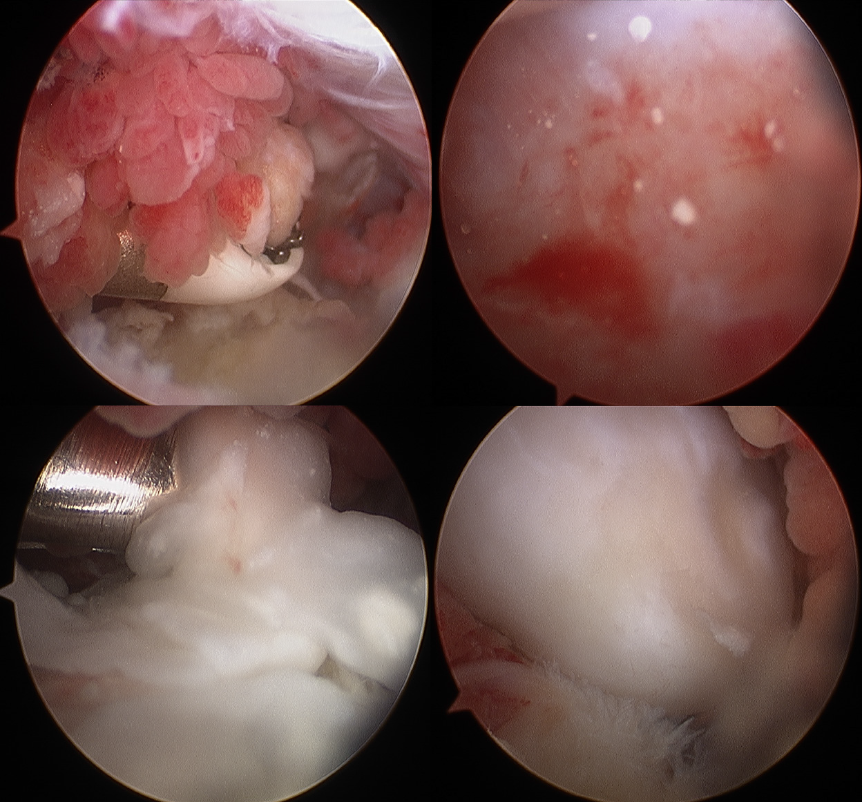
Figure 7
From the standard posterior portal, the arthroscopic view revealed a large number of cartilaginous loose bodies and synovial capsule hyperplasia, with inflammatory changes in the region of the subscapularis.
- Citation: Tang XF, Qin YG, Shen XY, Chen B, Li YZ. Arthroscopic surgery for synovial chondroma of the subacromial bursa with non-traumatic shoulder subluxation complications: Two case reports. World J Clin Cases 2022; 10(5): 1645-1653
- URL: https://www.wjgnet.com/2307-8960/full/v10/i5/1645.htm
- DOI: https://dx.doi.org/10.12998/wjcc.v10.i5.1645


