Copyright
©The Author(s) 2022.
World J Clin Cases. Feb 16, 2022; 10(5): 1639-1644
Published online Feb 16, 2022. doi: 10.12998/wjcc.v10.i5.1639
Published online Feb 16, 2022. doi: 10.12998/wjcc.v10.i5.1639
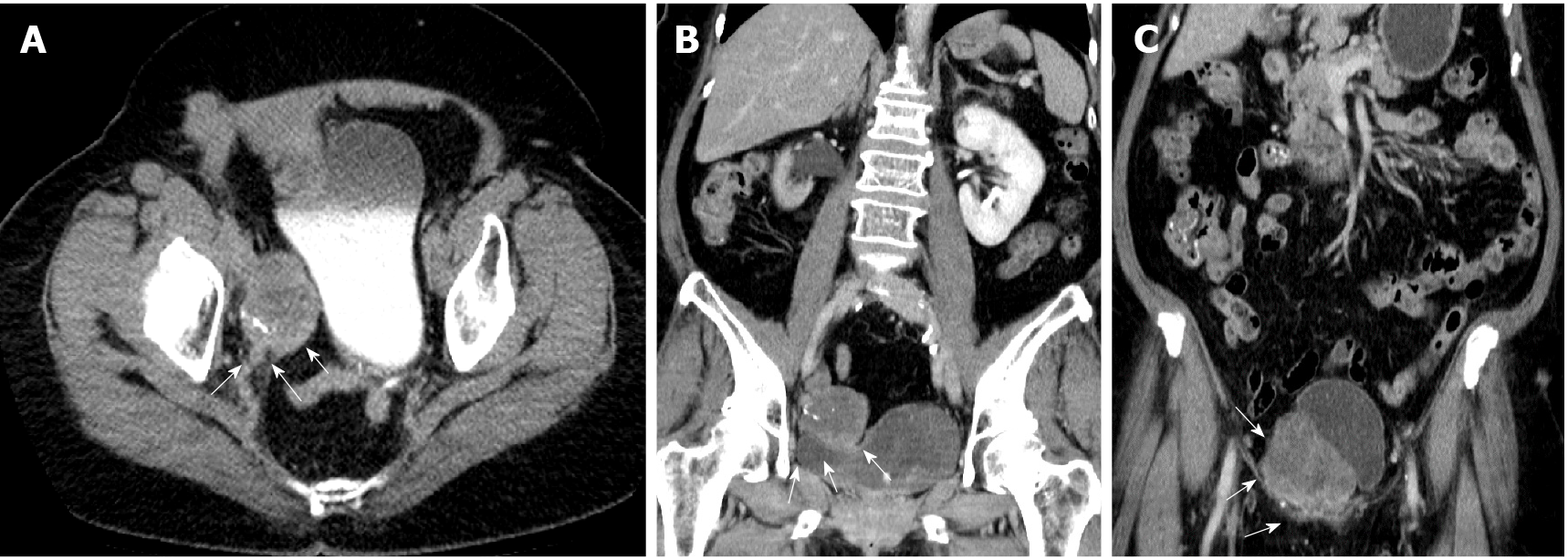
Figure 1 Abdominal computed tomography demonstrated that the tumor extended from the right distal third ureter to the ureterovesical junction.
A: White arrowhead in the axial view; B and C: Coronal view.
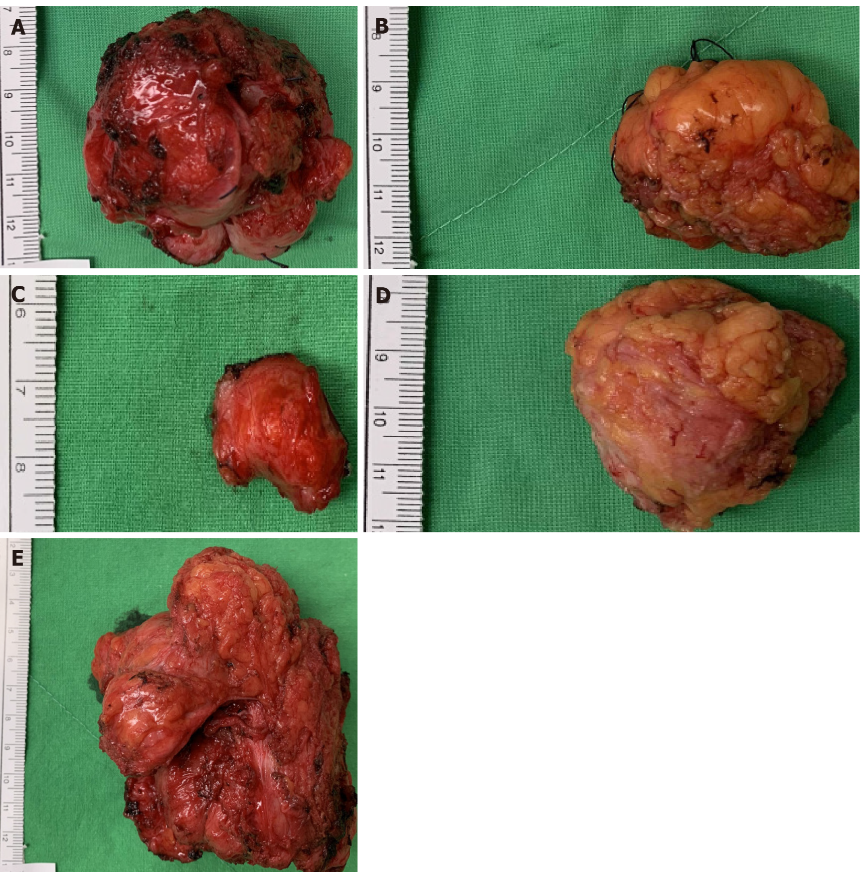
Figure 2 Intraoperative images of firm gray-whitish tumors.
A: Located at right distal ureter; B: Located at right inguinal area; C: Located at left inguinal area; D: Located at abdominal wall; E: Located at sigmoid colon.
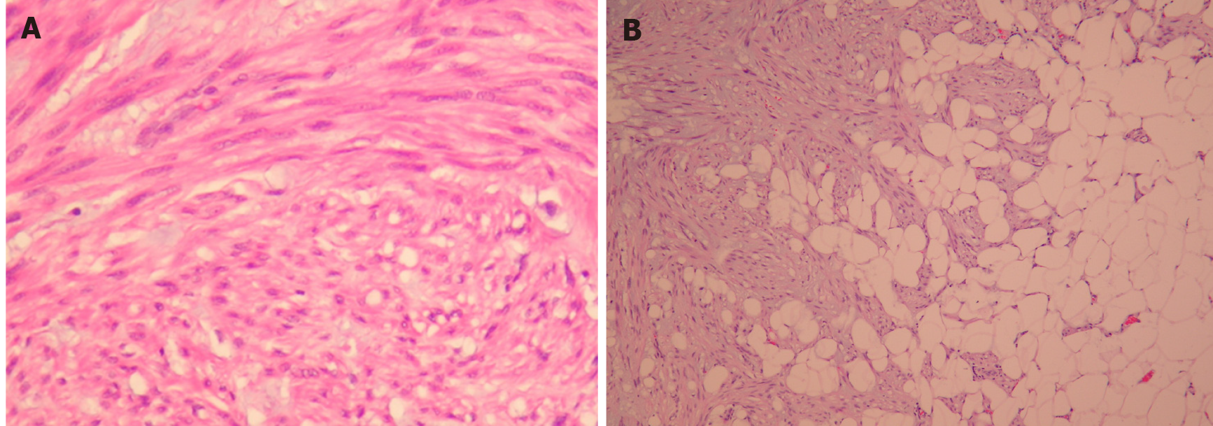
Figure 3 Histological appearance of disseminated peritoneal leiomyomatosis tumor.
A: The tumor has hypercellular areas with focal myxoid matrix, composed of spindled-shape neoplastic cells arranged in interlacing fascicles and storiform growth pattern (original magnification, 200 ×); B: Myxoid matrix infiltrated into adipose tissue (original magnification, 100 ×).
- Citation: Wen CY, Lee HS, Lin JT, Yu CC. Disseminated peritoneal leiomyomatosis with malignant transformation involving right ureter: A case report. World J Clin Cases 2022; 10(5): 1639-1644
- URL: https://www.wjgnet.com/2307-8960/full/v10/i5/1639.htm
- DOI: https://dx.doi.org/10.12998/wjcc.v10.i5.1639


