Copyright
©The Author(s) 2022.
World J Clin Cases. Feb 16, 2022; 10(5): 1617-1622
Published online Feb 16, 2022. doi: 10.12998/wjcc.v10.i5.1617
Published online Feb 16, 2022. doi: 10.12998/wjcc.v10.i5.1617
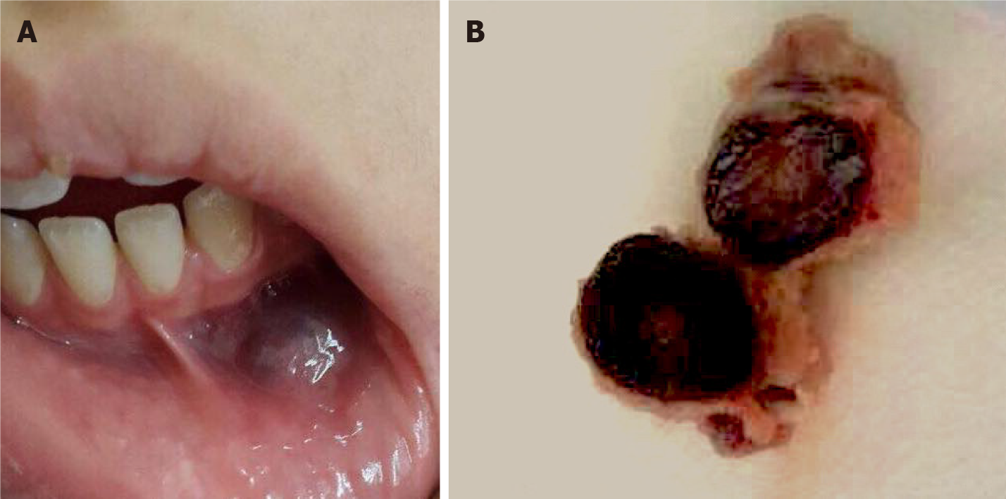
Figure 1 Intraoral examination and gross examination of the surgical specimen.
A: Intraoral lesion measuring 2.0-1.5 cm, solitary, fluctuant, bluish, smooth, palpable submucosal mass located in the left mandibular vestibule; B: A single, solid mass, bluish in color, and rubbery in consistency.
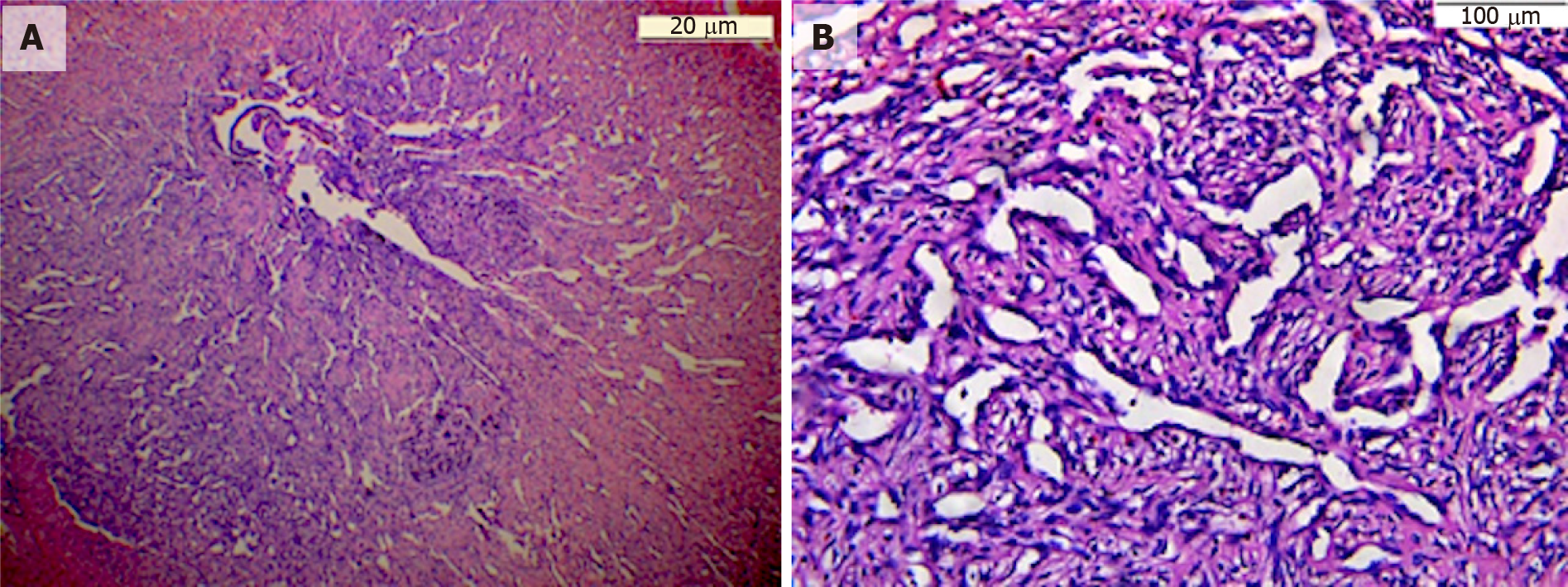
Figure 2 Histopathological analysis showed numerous thin-walled blood vessels with dilated blood vessel spaces, areas of hemorrhage and chronic inflammatory cell infiltrate which is consistent with capillary hemangioma in the labial vestibule.
A: 20 μm; B: 100 μm.
- Citation: Aloyouny AY, Alfaifi AJ, Aladhyani SM, Alshalan AA, Alfayadh HM, Salem HM. Hemangioma in the lower labial vestibule of an eleven-year-old girl: A case report. World J Clin Cases 2022; 10(5): 1617-1622
- URL: https://www.wjgnet.com/2307-8960/full/v10/i5/1617.htm
- DOI: https://dx.doi.org/10.12998/wjcc.v10.i5.1617


