Copyright
©The Author(s) 2022.
World J Clin Cases. Feb 16, 2022; 10(5): 1602-1608
Published online Feb 16, 2022. doi: 10.12998/wjcc.v10.i5.1602
Published online Feb 16, 2022. doi: 10.12998/wjcc.v10.i5.1602
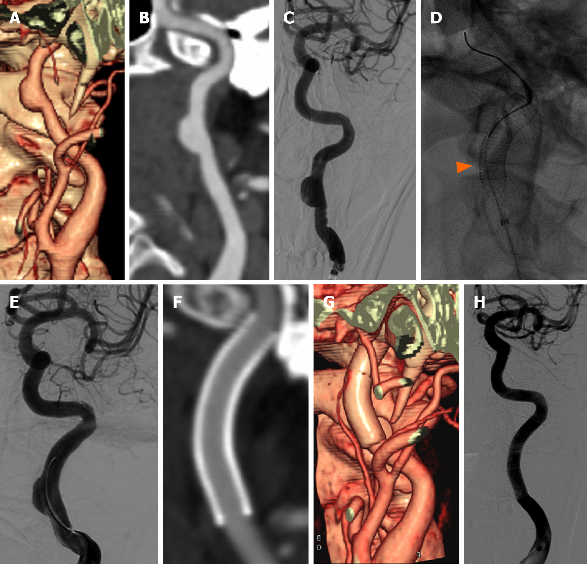
Figure 1 Case 1 was a 57-year-old male patient with sudden right limb weakness and vague speech and was diagnosed with cerebral infarction in February 2019.
He was ultimately treated using a SUPERA stent. A: Cervical computed tomographic angiography (CTA) volume reconstruction; B: curved surface reconstruction show that the wide-necked dissecting aneurysm was situated on the upper segment of the left internal carotid artery; C: confirmed by digital subtraction angiography (DSA); D and E: The patient underwent SUPERA stent endovascular therapy, the arrow shows the dense stent mesh; F and G: Three months later, cervical CTA showed that the aneurysm had disappeared completely. H: One year later, DSA showed that the internal carotid artery was repaired perfectly.
Figure 2 Case 2 was a 57-year-old man who suddenly felt dizzy and developed unsteady walking in November 2019.
He was ultimately treated using a SUPERA stents. A: Cervical computed tomographic angiography (CTA) volume reconstruction; B: digital subtraction angiography (DSA) showed a fusiform dilated dissecting aneurysm with severe stenosis located in the upper segment of the right internal carotid artery, involving the petrous segment; C and D: It was treated with SUPERA stent endovascular treatment, the arrow shows the dense stent reticular wire; E: Three months later, cervical CTA showed that the aneurysm had disappeared completely, and the lumen of the internal carotid artery was unobstructed; F: One year later, DSA showed that the internal carotid artery was repaired perfectly.
- Citation: Qiu MJ, Zhang BR, Song SJ. Treatment of extracranial internal carotid artery dissecting aneurysm with SUPERA stent implantation: Two case reports. World J Clin Cases 2022; 10(5): 1602-1608
- URL: https://www.wjgnet.com/2307-8960/full/v10/i5/1602.htm
- DOI: https://dx.doi.org/10.12998/wjcc.v10.i5.1602


