Copyright
©The Author(s) 2022.
World J Clin Cases. Feb 16, 2022; 10(5): 1586-1591
Published online Feb 16, 2022. doi: 10.12998/wjcc.v10.i5.1586
Published online Feb 16, 2022. doi: 10.12998/wjcc.v10.i5.1586
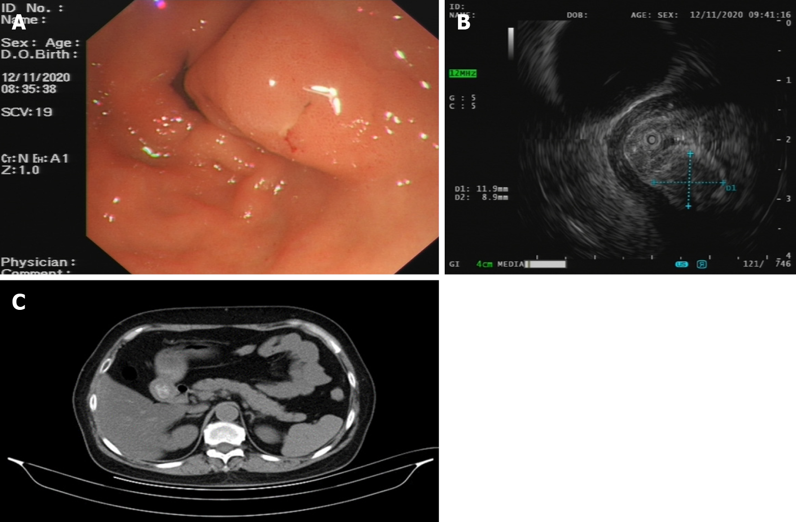
Figure 1 Preoperative imaging examinations.
A: Gastroscopy revealing a submucosal protuberance of the gastric antrum; B: Endoscopic ultrasonography indicating a round mixed echogenic mass with a size of approximately 1.19 cm × 0.89 cm; C: Abdominal computed tomography scan showing no obvious abnormal thickening or enhancement shadow of the gastric antrum.
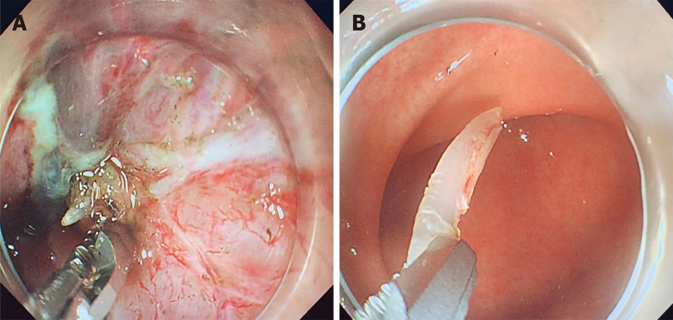
Figure 2 Surgical images.
A: During the endoscopic submucosal dissection, a white strip of a foreign body was found under the wound; B: A fish bone approximately 20 mm in length was removed using a dual knife.
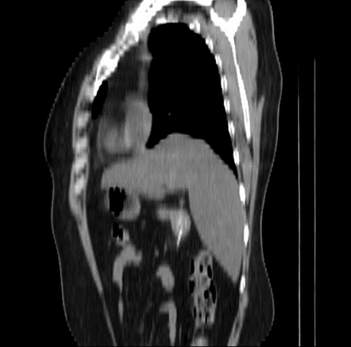
Figure 3 Computed tomography reconstruction of a fishbone-like (approximately 20 mm long) high-density image.
- Citation: Du WW, Huang T, Yang GD, Zhang J, Chen J, Wang YB. Submucosal protuberance caused by a fish bone in the absence of preoperative positive signs: A case report. World J Clin Cases 2022; 10(5): 1586-1591
- URL: https://www.wjgnet.com/2307-8960/full/v10/i5/1586.htm
- DOI: https://dx.doi.org/10.12998/wjcc.v10.i5.1586


