Copyright
©The Author(s) 2022.
World J Clin Cases. Feb 6, 2022; 10(4): 1333-1340
Published online Feb 6, 2022. doi: 10.12998/wjcc.v10.i4.1333
Published online Feb 6, 2022. doi: 10.12998/wjcc.v10.i4.1333
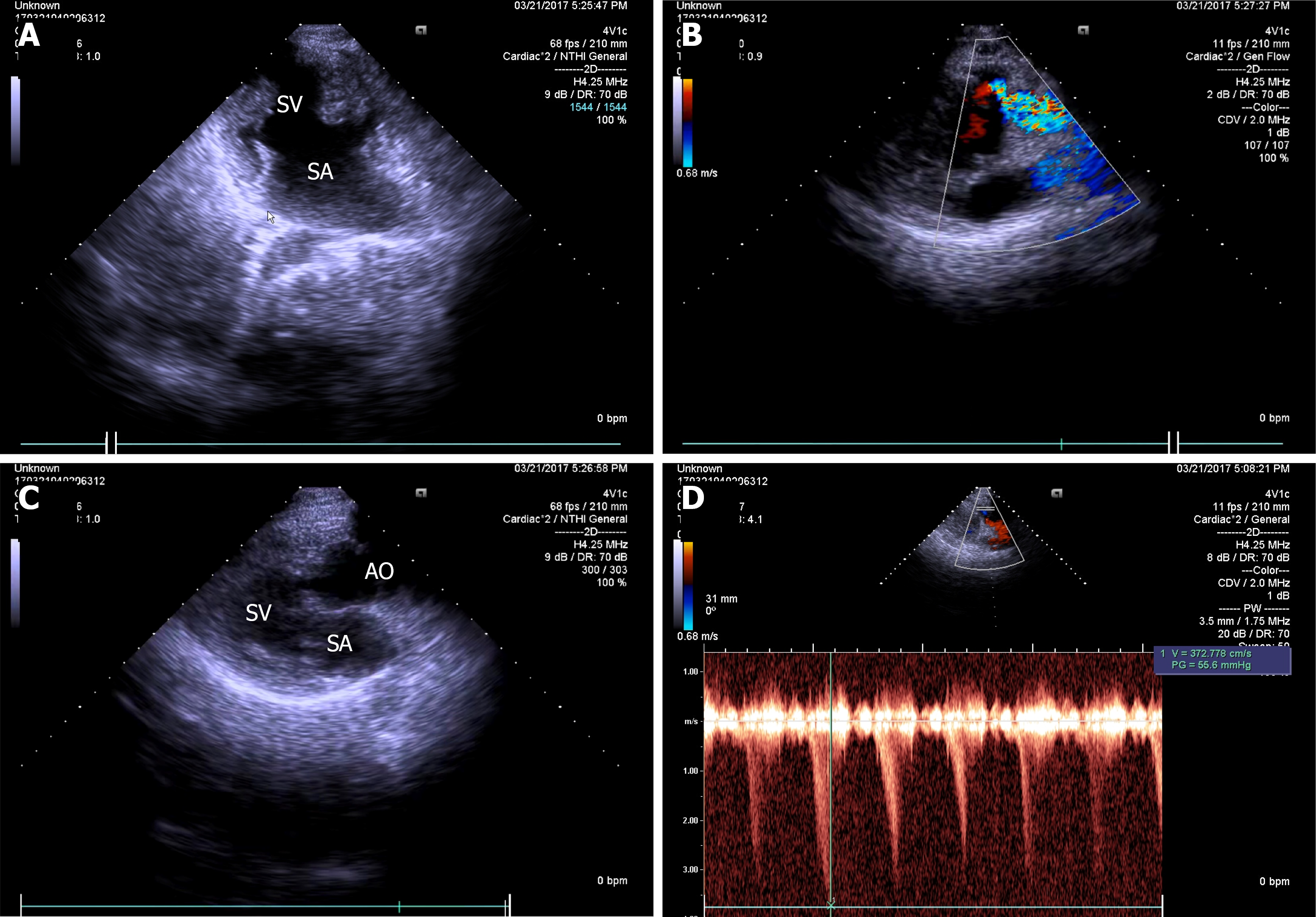
Figure 1 Two-dimensional echocardiographic apical 4-chamber view.
A: Common atrium, single ventricle; B: Hypoplastic pulmonary artery arising from single ventricle; C: Aorta arising from single ventricle; D: Continuous wave Doppler at pulmonary valve with gradient of 55.6 mmHg. SV: Single ventricle; SA: Single atrium; AO: Aorta; PA: Pulmonary artery.
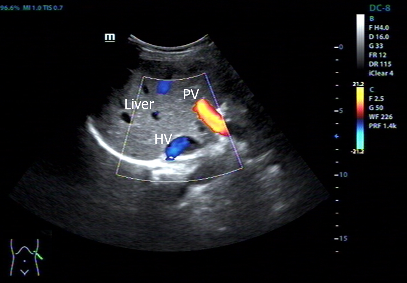
Figure 2 Abdominal B-ultrasound showing the liver was on the left side and the spleen on the right.
PV: Splenic vein; HV: Hepatic vein.
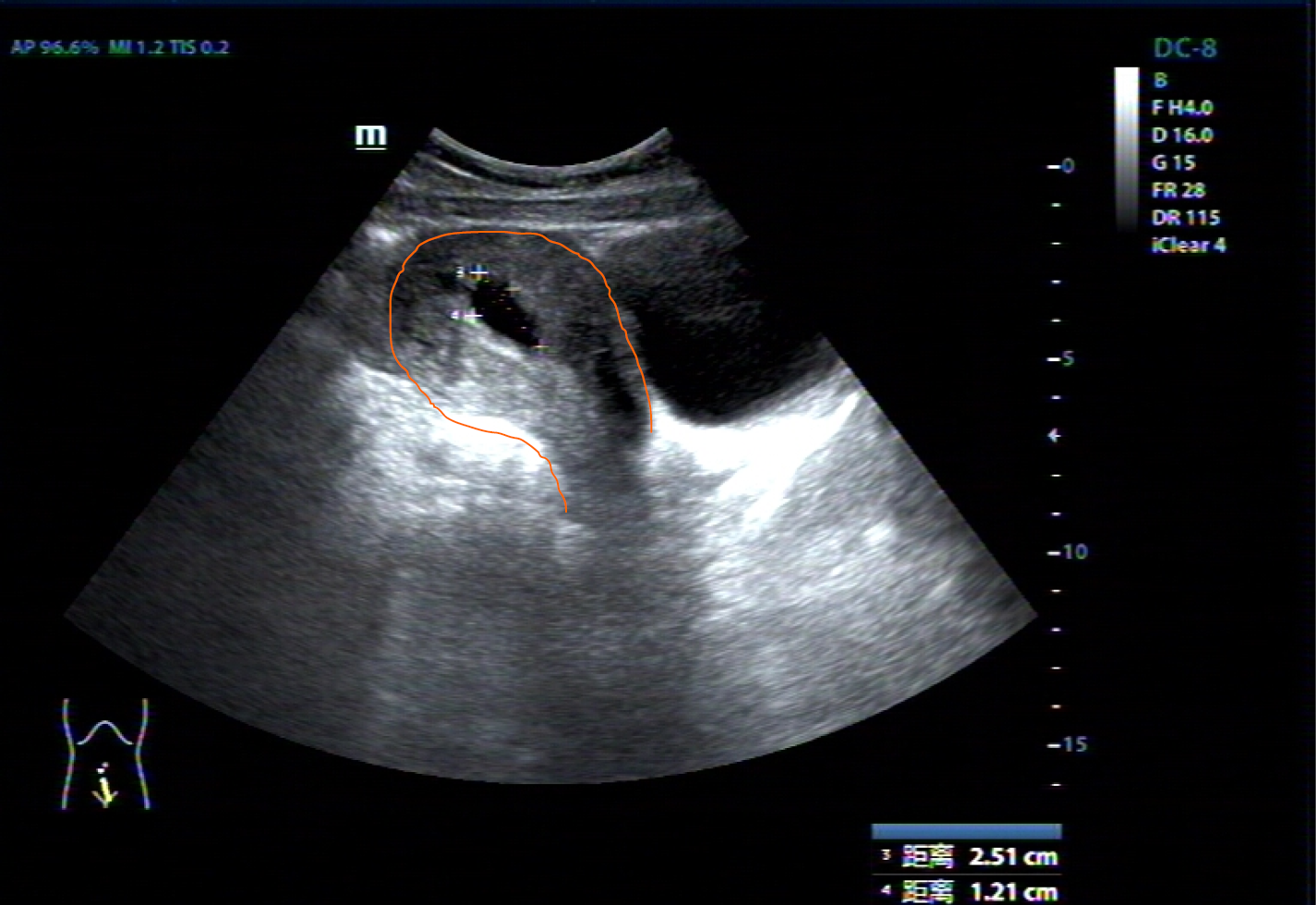
Figure 3 Abdominal B-ultrasound findings.
The angle between the cervix and the uterine body is located behind the uterus. The uterus has left and right mirror inversion. BL: Bladder; UT: Uterus; GS: Gestation sac.
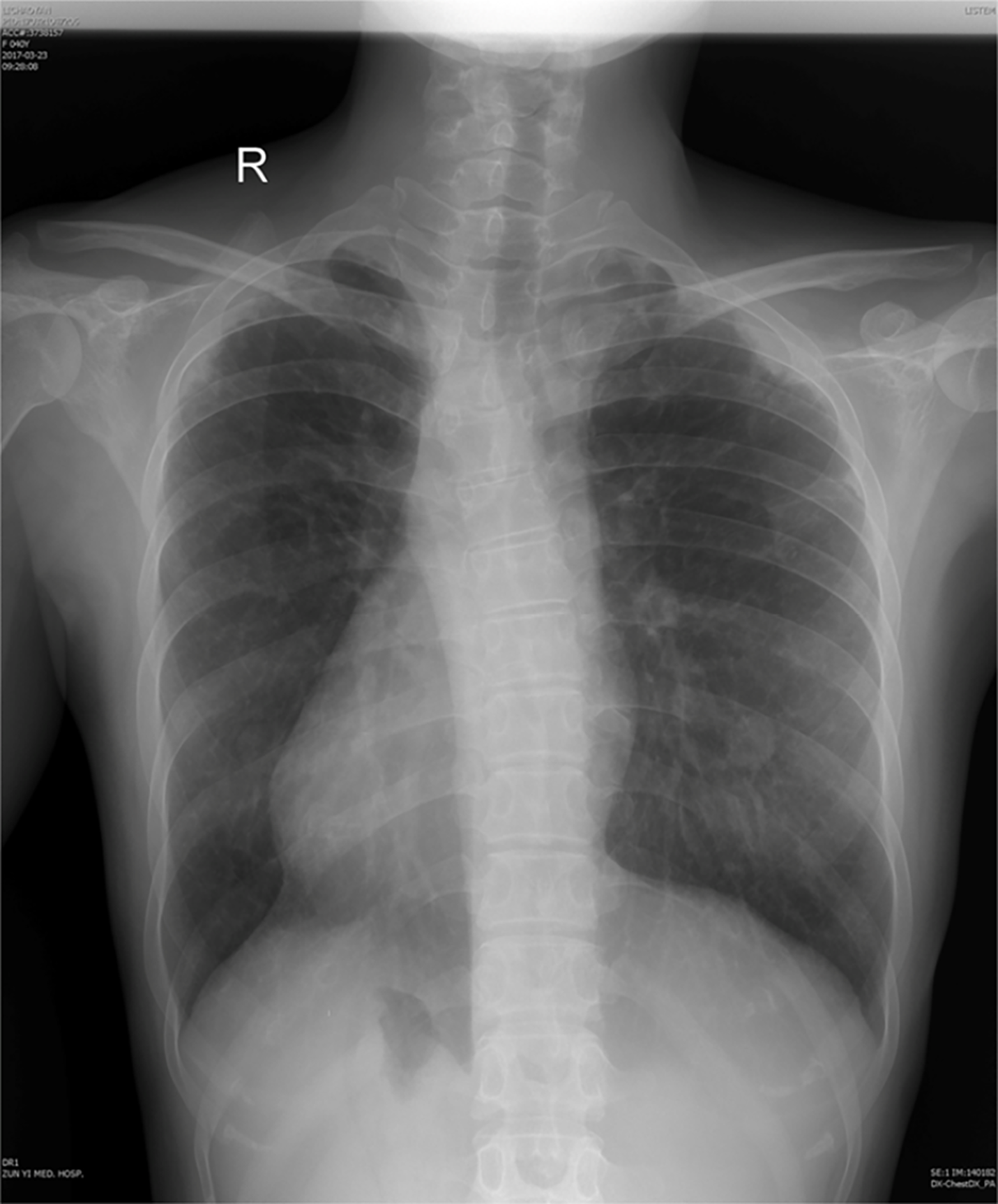
Figure 4 X-ray showing the heart is on the right side of the chest, with the apex to the right.
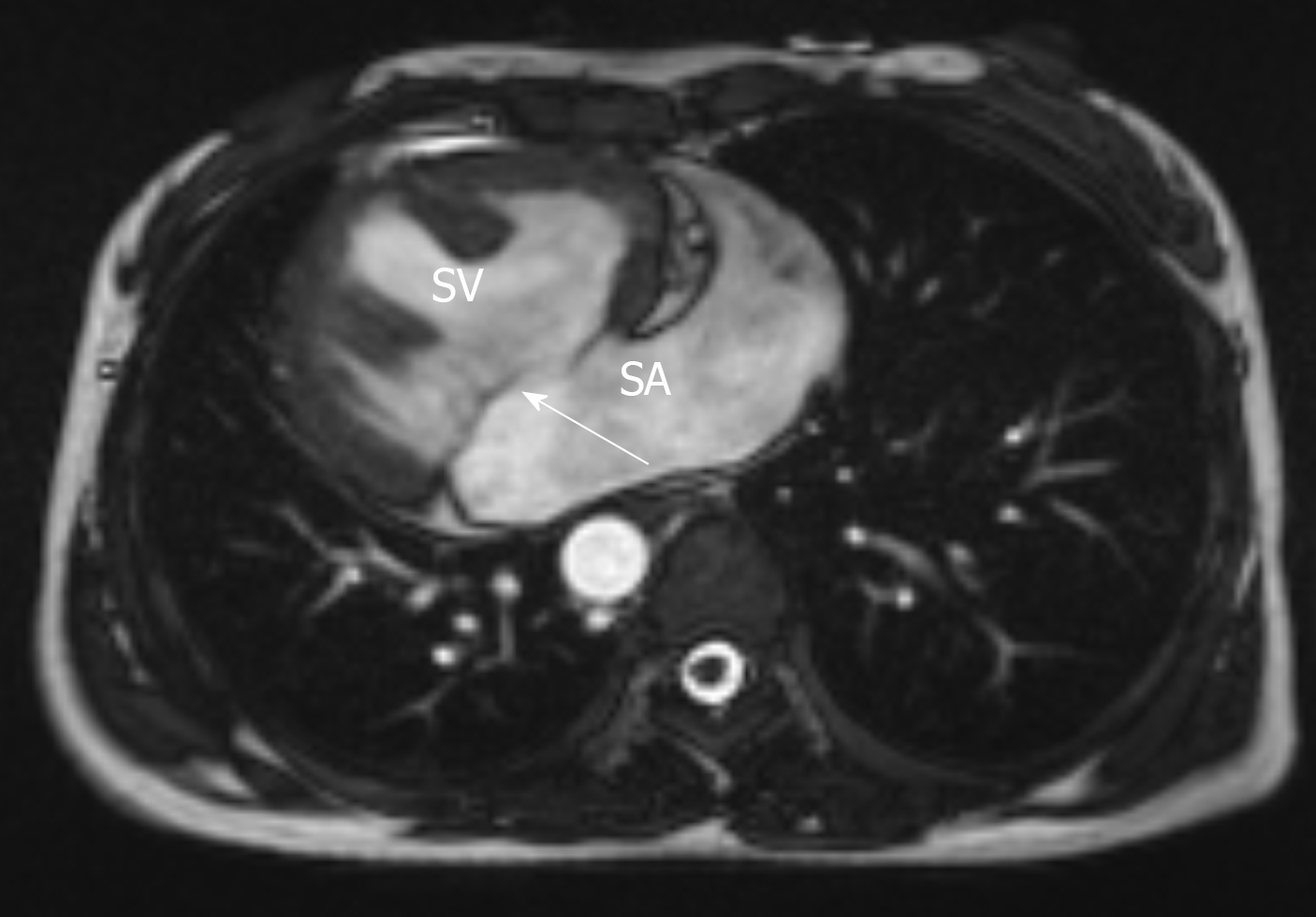
Figure 5 Cardiac magnetic resonance imaging showing SV with anatomical left ventricular morphology, SA and common atrioventricular valve (arrow).
SV: Single ventricle; SA: Single atrium.
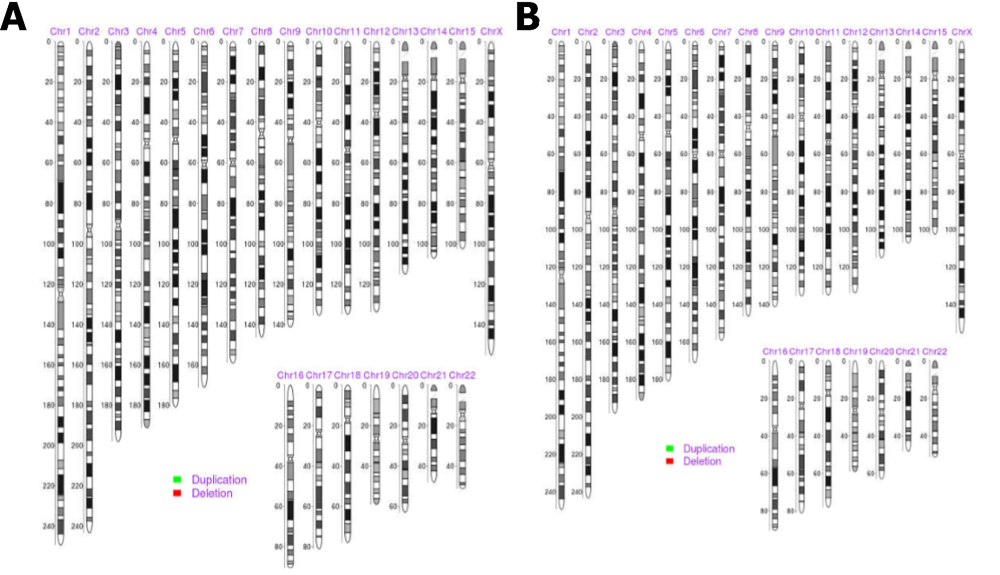
Figure 6 Chromosome analysis.
A: Peripheral blood; B: Villus tissues, showing no suppressive, pathogenic, and clear chromosomal microdeletion/ microduplication syndrome or aneuploidy abnormality, based on the GRCH37/hg19 N reference genome, representing the X or Y chromosome.
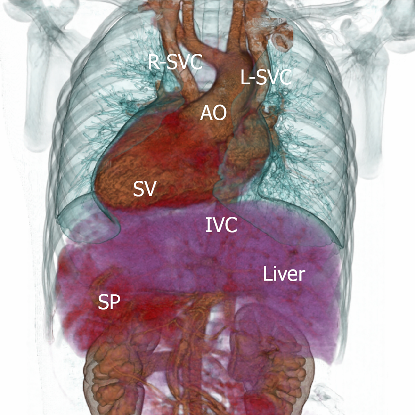
Figure 7 Patient’s viscera distribution sketch: single ventricle is located on the right side of thoracic cavity, apex of heart points to the right lower side.
Aorta position is anterior to SV, superior vena cava is divided into two and converges into SA. The liver is located in the left upper abdomen, the spleen is located in the right upper abdomen, and the inferior vena cava is located in the left side of the midline. R-SVC: Right superior vena cava; L-SVC: Left superior vena cava; SV: Single ventricle; AO: Aorta; IVC: Inferior vena cava; SP: Spleen.
- Citation: Duan HZ, Liu JJ, Zhang XJ, Zhang J, Yu AY. Multiple miscarriages in a female patient with two-chambered heart and situs inversus totalis: A case report . World J Clin Cases 2022; 10(4): 1333-1340
- URL: https://www.wjgnet.com/2307-8960/full/v10/i4/1333.htm
- DOI: https://dx.doi.org/10.12998/wjcc.v10.i4.1333


