Copyright
©The Author(s) 2022.
World J Clin Cases. Dec 16, 2022; 10(35): 12880-12889
Published online Dec 16, 2022. doi: 10.12998/wjcc.v10.i35.12880
Published online Dec 16, 2022. doi: 10.12998/wjcc.v10.i35.12880
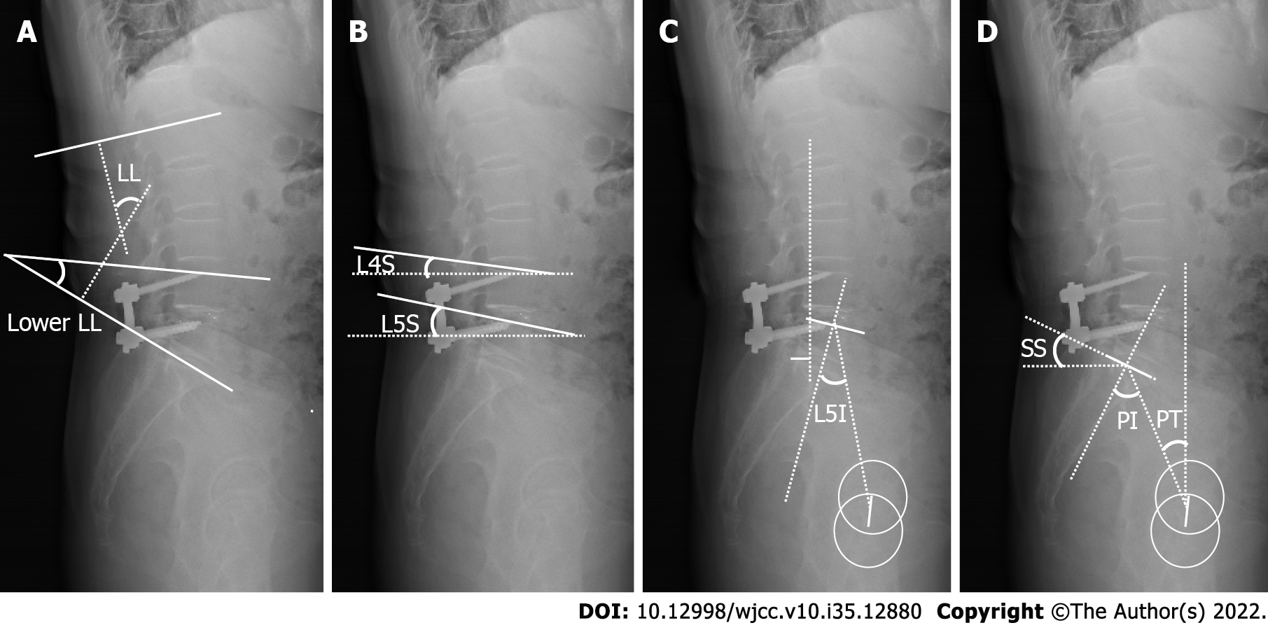
Figure 1 Measurement of sagittal lumbar-pelvic parameters.
A: Lumbar lordosis (LL), the angle between the upper endplate of L1 and the sacrum platform; Lower LL, the angle between the upper endplate of L4 and the sacral platform; B: L4 slope and L5 slope, the angle between the upper endplate of L4 or L5 and the horizontal line; C: L1 axis and S1 distance, the distance between the plumb line through the L1 vertebra center and the posterior upper edge of the sacrum platform; and L5 incidence; D: Pelvic incidence, the angle between the perpendicular of the upper endplate of S1 and the line joining the middle of the upper endplate of S1 and the hip axis (midway between the centers of the two femoral heads); pelvic tilt, the angle between the vertical line and the line joining the middle of the upper endplate of S1 and the hip axis; and sacral slope, the angle between the upper endplate of S1 and the horizontal line. LL: Lumbar lordosis; L4S: L4 slope; L5S: L5 slope; L5I: L5 incidence; SS: Sacral slope; PI: Pelvic incidence; PT: Pelvic tilt.
Figure 2 Sagittal lumbar-pelvic parameters of a typical case.
A 68-year-old female with complaints of lower back pain, right posterolateral lower-extremity numbness, and intermittent claudication for five months. Diagnosis: lumbar disc herniation, lumbar spinal stenosis (L4-5). Treatment: L4-5 midline lumbar fusion was performed, and postoperative symptoms were significantly improved. A: Preoperative sagittal magnetic resonance imaging; B: Preoperative anteroposterior and lateral spine X-ray; C: Anteroposterior and lateral lumbar spine X-ray at the final follow-up.
- Citation: Wang YT, Li BX, Wang SJ, Li CD, Sun HL. Radiological and clinical outcomes of midline lumbar fusion on sagittal lumbar-pelvic parameters for degenerative lumbar diseases. World J Clin Cases 2022; 10(35): 12880-12889
- URL: https://www.wjgnet.com/2307-8960/full/v10/i35/12880.htm
- DOI: https://dx.doi.org/10.12998/wjcc.v10.i35.12880


