Copyright
©The Author(s) 2022.
World J Clin Cases. Dec 16, 2022; 10(35): 12854-12874
Published online Dec 16, 2022. doi: 10.12998/wjcc.v10.i35.12854
Published online Dec 16, 2022. doi: 10.12998/wjcc.v10.i35.12854
Figure 1 Anembryonic pregnancy.
30-year-old, first pregnancy, complaining of vaginal bleeding. Ultrasonography revealed a gestational sac, slightly irregular border with a mean sac diameter of 28 mm (thick arrow). There was no yolk sac or embryo in the sac. There was also minimal perigestational hemorrhage in the superolateral of the sac (thin arrows).
Figure 2 Lower Uterine Segment Gestation.
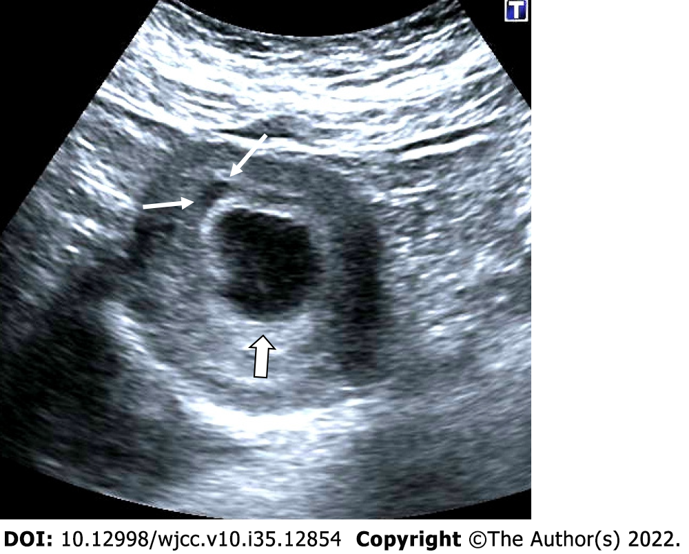
25-year-old, first pregnancy. On ultrasonography, a 6-week well-circumscribed gestational sac is seen low located close to the cervix (Thick arrows: Cervix; thin arrow: Gestational sac).
Figure 3 Complete miscarriage.
A: 26-year-old, first pregnancy, 6 wk pregnant by date of the last menstrual period and complaining of vaginal bleeding. No gestational sac or fetal structure was seen in the endometrial cavity on axial section ultrasonography; There was enlargement in the cervical canal and hypoechoic collection at this level; B: Coronal section; C: Sagittal section, thin arrows. On ultrasonography performed 3 days ago, a gestational sac was seen in the uterine cavity (not shown in the figure).
Figure 4 Subamniotic and retroplacental hemorrhage.
A and B: Retroplacental hemorrhage. 18 wk pregnant, complaining of vaginal bleeding. On ultrasonography, there was a hypoechoic collection area (arrows) extending in a long segment posterior to the placenta (asterisk); C: Subamniotic hemorrhage, 12 wk pregnant, complaining of pain. On ultrasonography, there was a heterogeneous collection area (arrows) located anterior of the placenta, reaching 10 cm at its widest part (asterisk: Placenta); D: Subamniotic hemorrhage, 18 wk pregnant. On ultrasonography, there was a hypoechoic, minimally heterogeneous collection area (arrows) anterior to the placenta (asterisk: Placenta).
Figure 5 Intraamniotic hemorrhage.
A-C: 21 wk pregnant, on ultrasonography, there were diffuse moving echogenicities (arrows) filling the entire amniotic cavity.
Figure 6 Partial hydatiform mole.
27-year-old, third pregnancy, 7 wk pregnant by date of the last menstrual period, complaining of pain, beta HCG value greater than 200000. A and C: On ultrasonography, there was a heterogeneous, echogenic mass appearance-enlarged multicystic placenta (thin arrows) that filled the endometrial cavity to a large extent, containing multiple millimetric cystic areas; B: The gestational sac was irregular and contained millimetric yolk sac (thick arrow).
Figure 7 Chorioamniotic separation.
A-C: In 3 different pregnant women between 14th and 15th weeks, separation is seen in the chorion and amniotic membranes (arrows). In the follow-ups, fusion occurred and no persistence was observed in the chorioamniotic separation (not shown in the figure).
Figure 8 Cystic hygroma.
24-year-old, first pregnancy, 12 wk of gestation. A: On ultrasonography, there was an increase in nuchal translucency (thin arrows); B: cystic areas of cystic hygroma (thick arrows) in the fetal cervical region.
Figure 9 Thickened nuchal fold.
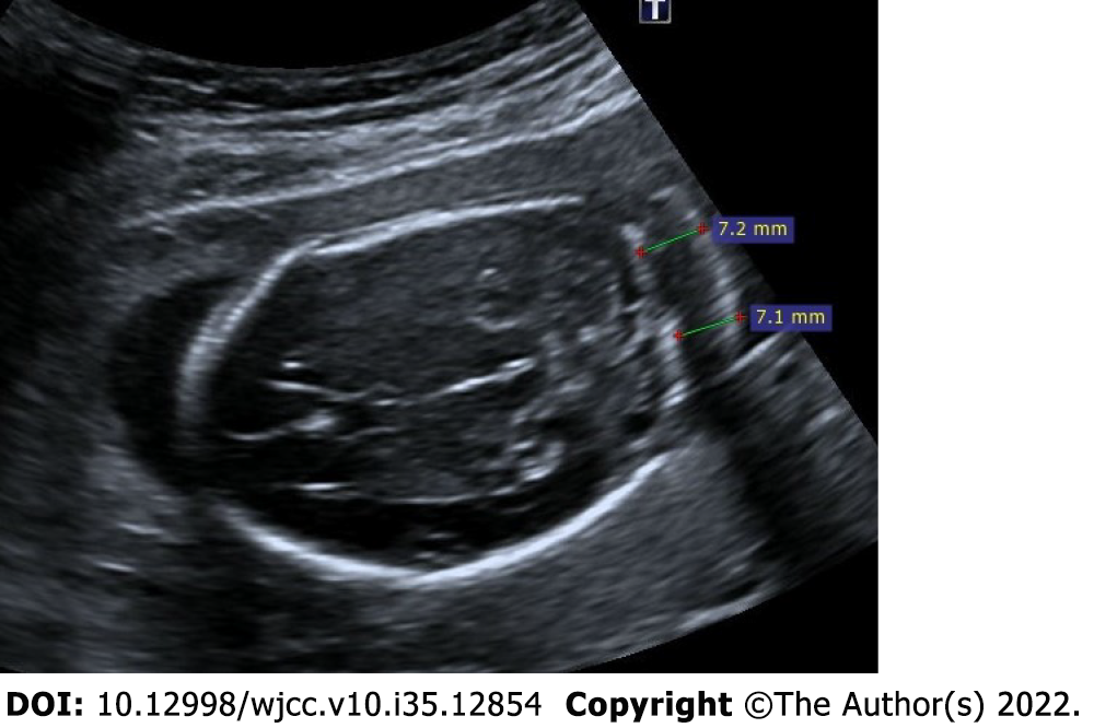
25-year-old, first pregnancy, 19 wk of gestation. On ultrasonography, there was an increase in the thickness of the nuchal fold measuring 7.2 mm.
Figure 10 Single umbilical artery.
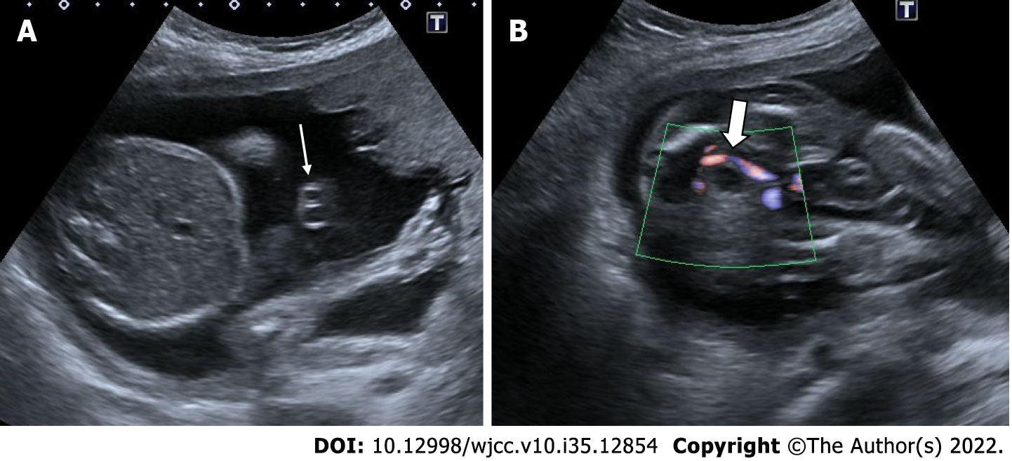
29-years-old, first pregnancy, 21 wk of gestation. A: On ultrasonography, there was one artery (thin arrow) and one vein in the umbilical cord; B: Doppler ultrasonography reveals a solitary artery at the level of the bladder in the fetal pelvis (thick arrow).
Figure 11 Acrania – exencephaly – anencephaly sequence.
21-year-old, pregnancy without follow-up, first examination. A-C: On ultrasonography, there was absence of fetal cranial bone structures (acrani) and exophytic extension of fetal cerebral tissue (exencephaly) (arrows).
Figure 12 Acrania – exencephaly – anencephaly sequence.
A and B: On ultrasonography, first case had absence of cranial bone structures (acrani) and cerebral tissue with exophytic extension (exencephaly) (arrows); C and D: There is another case had acrani and exencephaly (arrows) on ultrasonography.
Figure 13 Alobar holoprosencephaly.
30-year-old, 21 wk of gestation. A: On ultrasonography, supratentorial cerebral structures are absent (thin arrows); B: There was bilateral hypotelorism-anophthalmia and a single orbital structure with exophytic extension from the midline (cyclopi) (thick arrow); C and D: The same patient also had a major cardiac anomaly (circle).
Figure 14 Isolated bilateral mild ventriculomegaly.
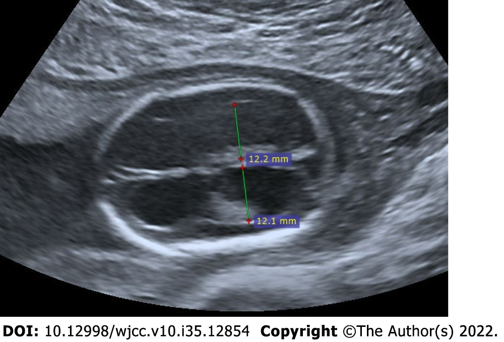
22-year-old, first pregnancy, 19 wk of gestation. On ultrasonography, the measurements of both ventricles were 12 mm and larger than normal.
Figure 15 Isolated choroid plexus cysts.
25-year-old, second pregnancy, 20 wk of gestation. A and B: On ultrasonography, there were anechoic choroid plexus cysts bilaterally reaching 1 cm diameter (diameters).
Figure 16 Myelomeningocele.
A: Ultrasonography showed an anechoic sac (diameters) extending outward from the posterior defect at the cervical level, reaching 3 cm in diameter; B: There is seen neural roots (arrows) within the sac.
Figure 17 Spinal dysraphism.
A-C: On ultrasonography, spina bifida was seen in the vertebral column at the lumbar-sacral level (arrows); D: There were lemon sign characterized by angulation in the bilateral frontoparietal suture (arrows); E: Additionaly, there were bilateral mild ventriculomegaly (diameters); F: There were banana sign (arrows) in the posterior fossa.
Figure 18 Occipital encephalocele.
30-year-old, second pregnancy, 20 wk of gestation. A-C: On ultrasonography, there was a herniated sac from the bone defect in the occipital region (thin arrows); C: A portion of the occipital lobe was present in the sac (thick arrow).
Figure 19 Megacystis.
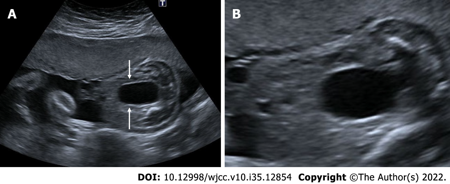
A: 25 wk of gestation. On ultrasonography, there was a distension reaching 4.5 cm in the bladder that did not regress in the follow-ups (arrows); B: Keyhole sign indicating an enlarged urethra is seen in the inferior of the bladder.
Figure 20 Bilateral mild hydronephrosis.
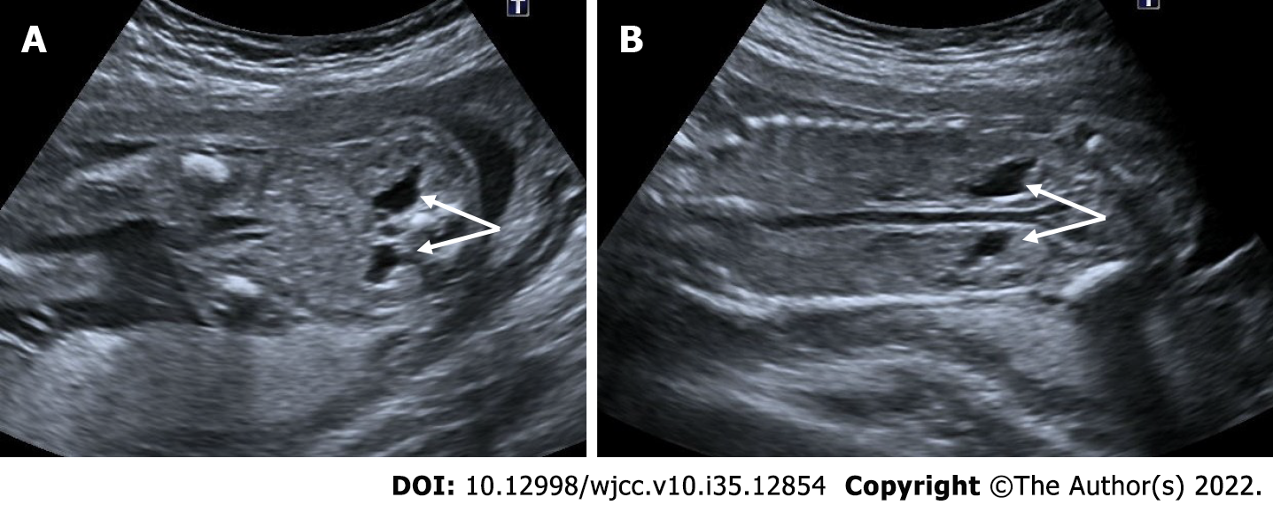
A and B: 23 wk of gestation. On ultrasonography, bilateral renal pelvis was wider than normal (arrows). Renal pelvis diameters were 6 mm on the right and 5 mm on the left. There was no dilatation of the bilateral ureters.
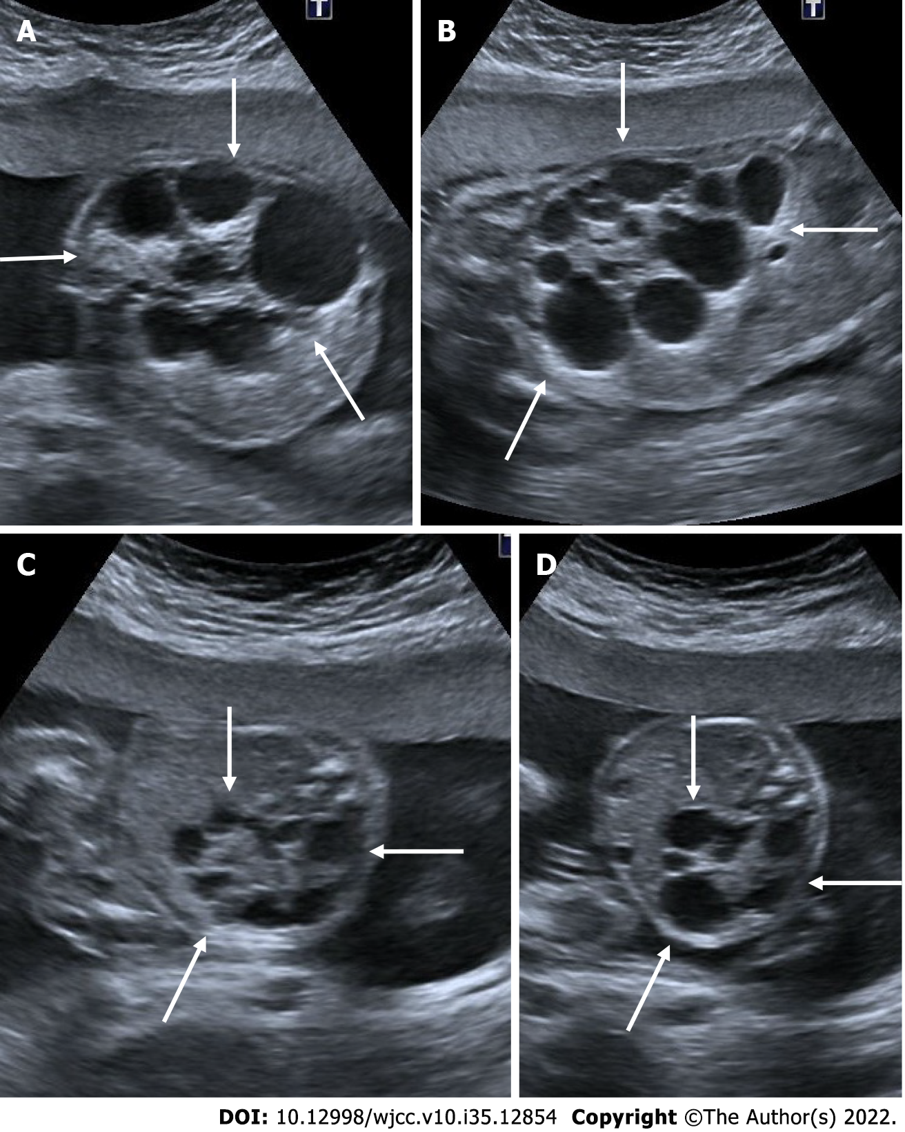
Figure 21 Multicystic dysplastic kidney.
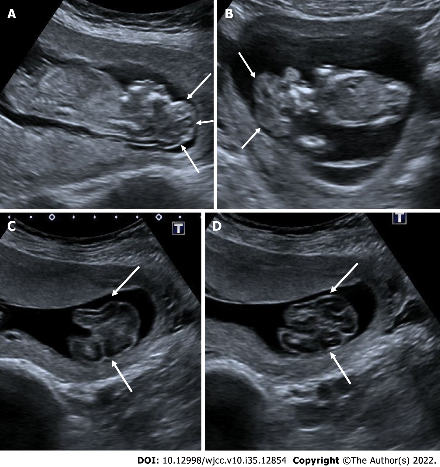
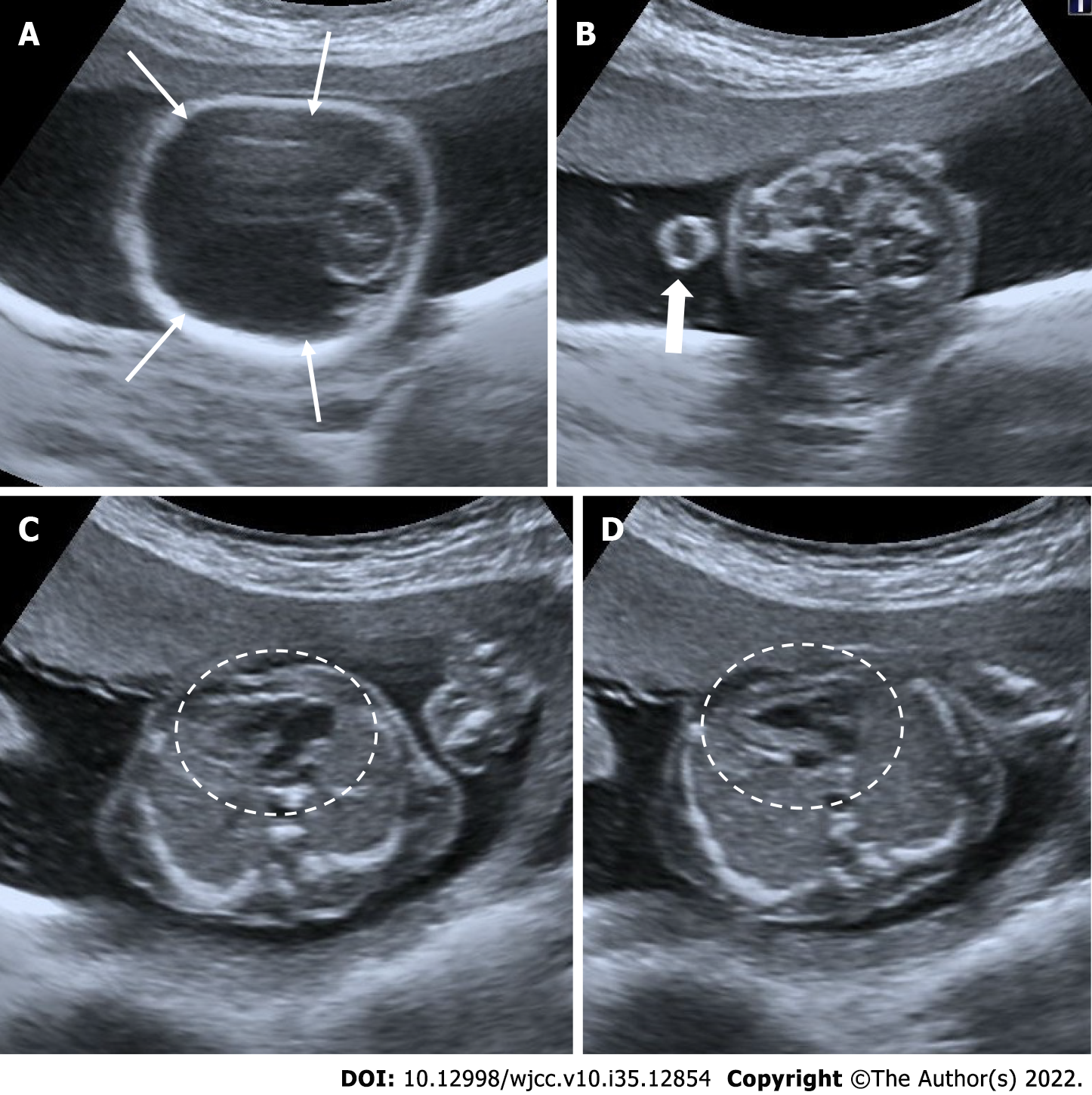
In two different pregnant women who underwent 2nd trimester obstetric sonography. A and B: Case 1, there was a multicystic appearance in different sizes with an increase in size in one kidney (arrows); C and D: Case 2; there was another case with a multicystic appearance in different sizes in one kidney (arrows).
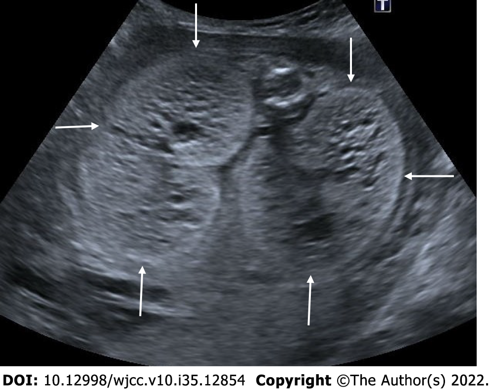
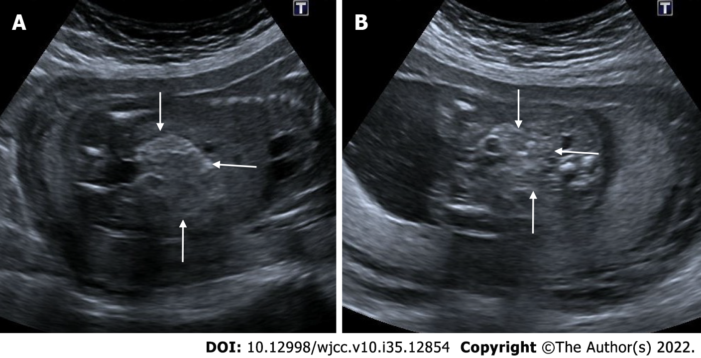
Figure 22 Autosomal recessive polycystic kidney disease.
27 wk of gestation. On ultrasonography, there was a significant increase in bilateral kidney sizes, multiple millimetric cystic heterogeneous echogenic kidneys (arrows). The patient also had oligohydramnios, cardiac and cranial anomalies (not shown in the figure).
Figure 23 Persistent right umbilical vein.
22 wk of gestation. A: On ultrasonography, while the stomach was in its normal position (asterisk); B and C: The umbilical vein (thin arrow) was seen to the right of the gallbladder (thick arrow).
Figure 24 Isolated cleft lip.
23 wk of gestation. A: Isolated cleft lip view in coronal section (arrow); B: Isolated cleft lip view in axial section (arrows).
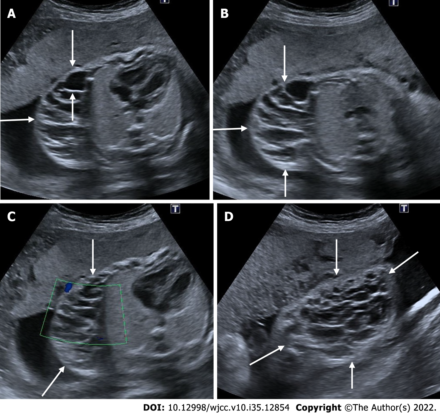
Figure 25 Cystic lymphangioma.
24 wk of gestation. A and B: On ultrasonography, a complex cystic heterogeneous lesion with multicystic areas on the right side of the fetal thoracic wall, under the skin (arrows); C: There was seen no vascularity on Doppler sonography; D: There is a view of the cystic lesion (arrows) on the chest wall from a different section.
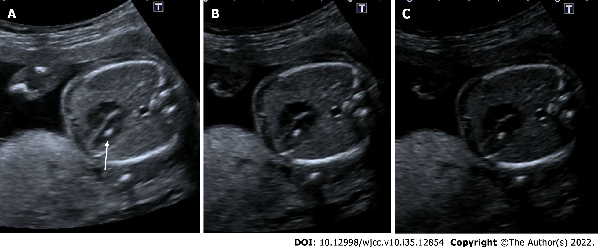
Figure 26 Echogenic intracardiac focus.
21 wk of gestation. A: On ultrasonography, there was one echogenic focus (arrow) in the left ventricle of the fetal heart; B and C: Although the gain settings were decreased, the hyperechoic appearance persisted.
Figure 27 Amniotic band sequence.
16 wk of gestation. A: On ultrasonography, there was amniotic band formation (thin arrows) in the right half of the amniotic cavity; B-D: Fetal cranium (thick arrows) was fixed in the secondary cavity formed at this level, and a deformed appearance was noted in the fetal cranium.
Figure 28 Echogenic fetal bowel.
22 wk of gestation. A and B: On ultrasonography, the intestines showed a diffuse echogenicity increase (arrows). Although intestinal echogenicity was not as echogenic as the bone structures in the same section, this case was later diagnosed as prenatal trisomy 21.
Figure 29 Omphalocele.
24 wk of gestation. A: On ultrasonography, abdominal wall defect and herniation at the level of umbilical cord insertion (thin arrows) are observed and the presence of intra-abdominal tissues and gall bladder (thick arrow) was noted within the herniation; B: The level of herniation is from the side of the umbilical cord, and the umbilical cord is seen in color Doppler; C: There is seen sagittal section view of the herniation.
- Citation: Ece B, Aydın S, Kantarci M. Antenatal imaging: A pictorial review. World J Clin Cases 2022; 10(35): 12854-12874
- URL: https://www.wjgnet.com/2307-8960/full/v10/i35/12854.htm
- DOI: https://dx.doi.org/10.12998/wjcc.v10.i35.12854


