Copyright
©The Author(s) 2022.
World J Clin Cases. Dec 6, 2022; 10(34): 12750-12760
Published online Dec 6, 2022. doi: 10.12998/wjcc.v10.i34.12750
Published online Dec 6, 2022. doi: 10.12998/wjcc.v10.i34.12750
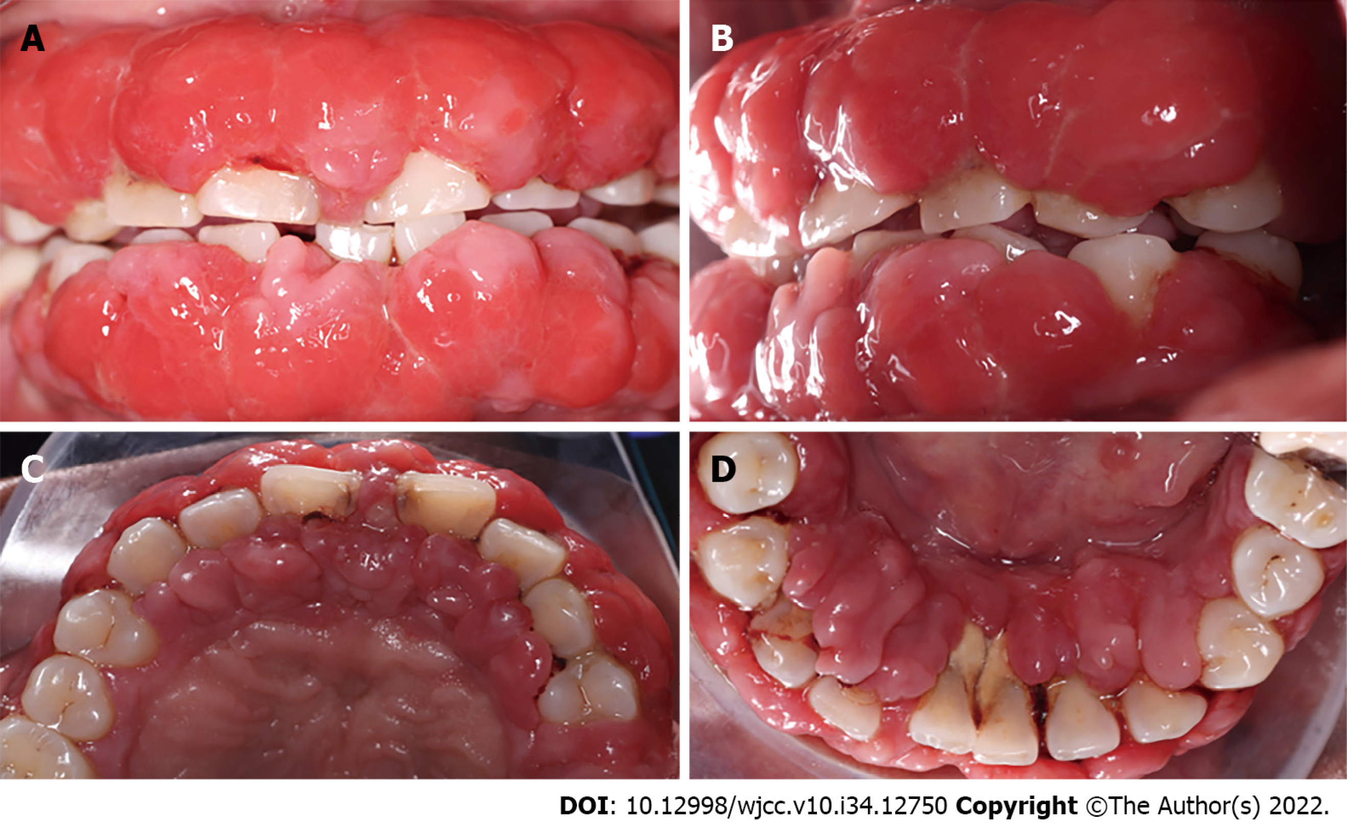
Figure 1 Gingival enlargement in the adult patient treated with cyclosporine.
In general, severe inflammation, redness of tissue, nodules, and calculus deposits were observed. A: Inter-occlusal view; B: Left side view; C: Upper occlusal view; D: Lower occlusal view.
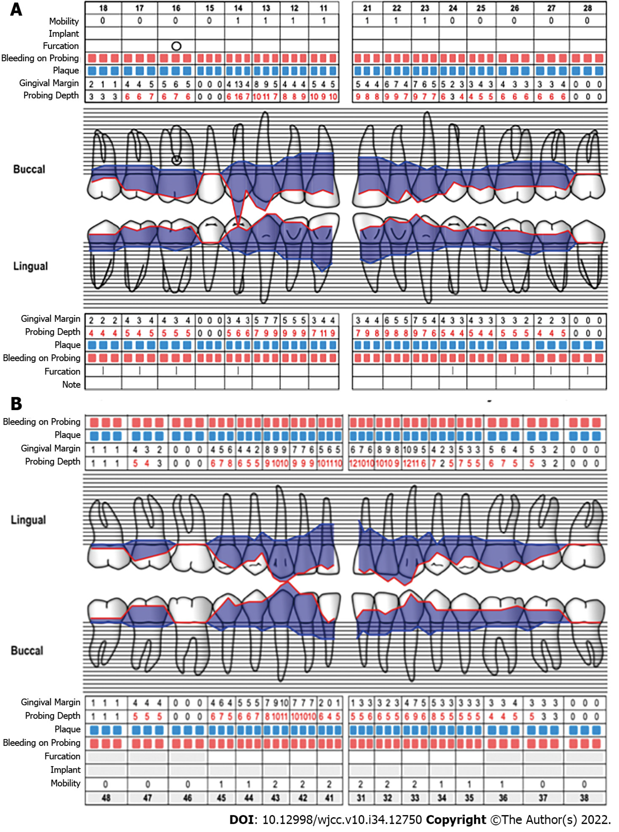
Figure 2 Periodontograms.
A: Upper; B: Lower.
Figure 3 Images from complete periapical study.
Generalized bone loss is evident, with greater loss in the incisors. A: Upper; B: Lower.
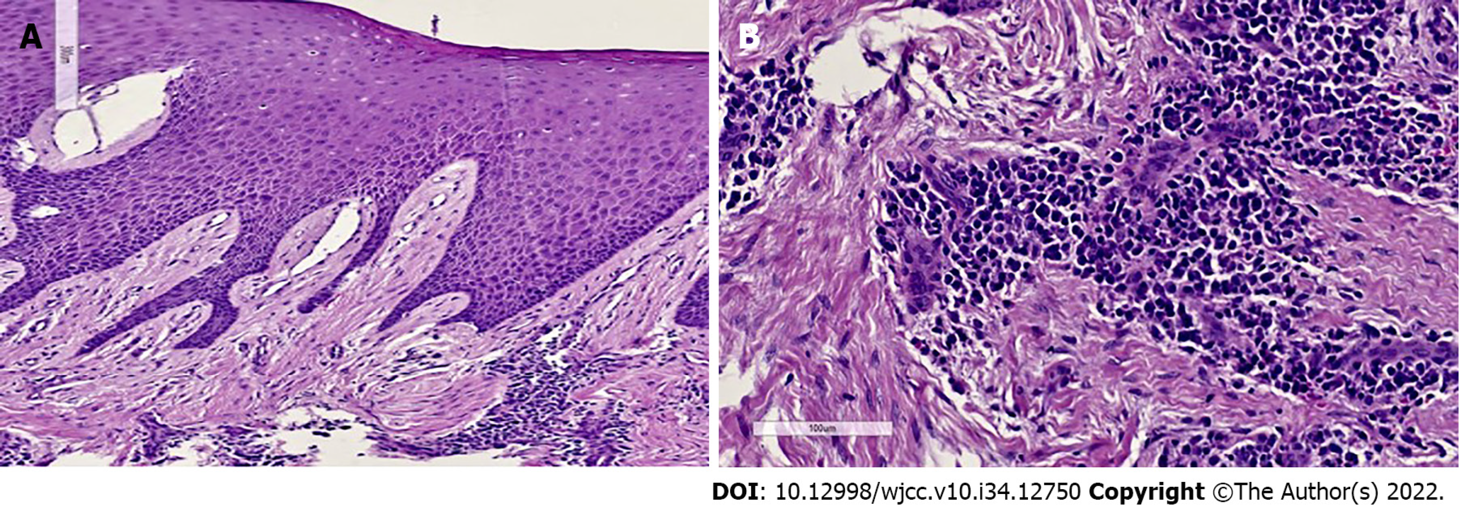
Figure 4 Gingival histopathology.
A: Fibroconnective tissue growth, elongated epithelial nails, and pseudo-epithelionomatous hyperplasia; B: Connective tissue showing large focal areas of chronic inflammatory infiltration and lymphoplasmacytic dispersion in a stroma of collagen fibers and active fibroblasts.
Figure 5 Tissue conditions 3 mo after initial phase.
The features depict decreased inflammation, pink coloration in interproximal areas, and smaller nodules. A: Inter-occlusal view; B: Upper sextant.
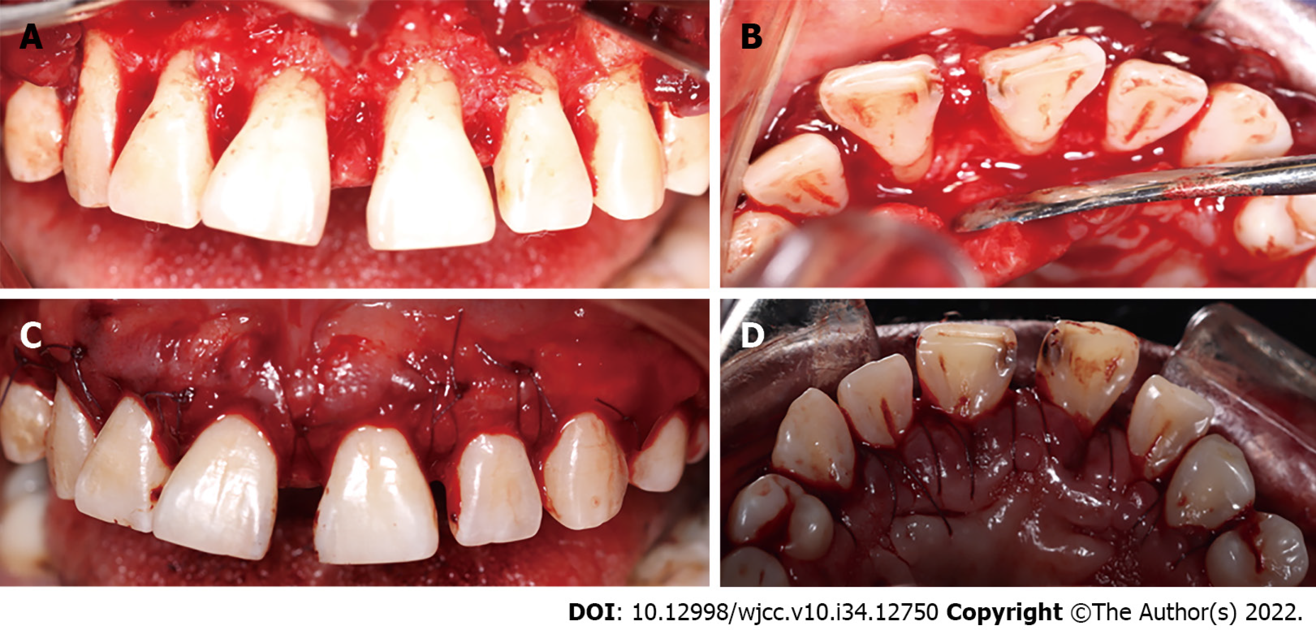
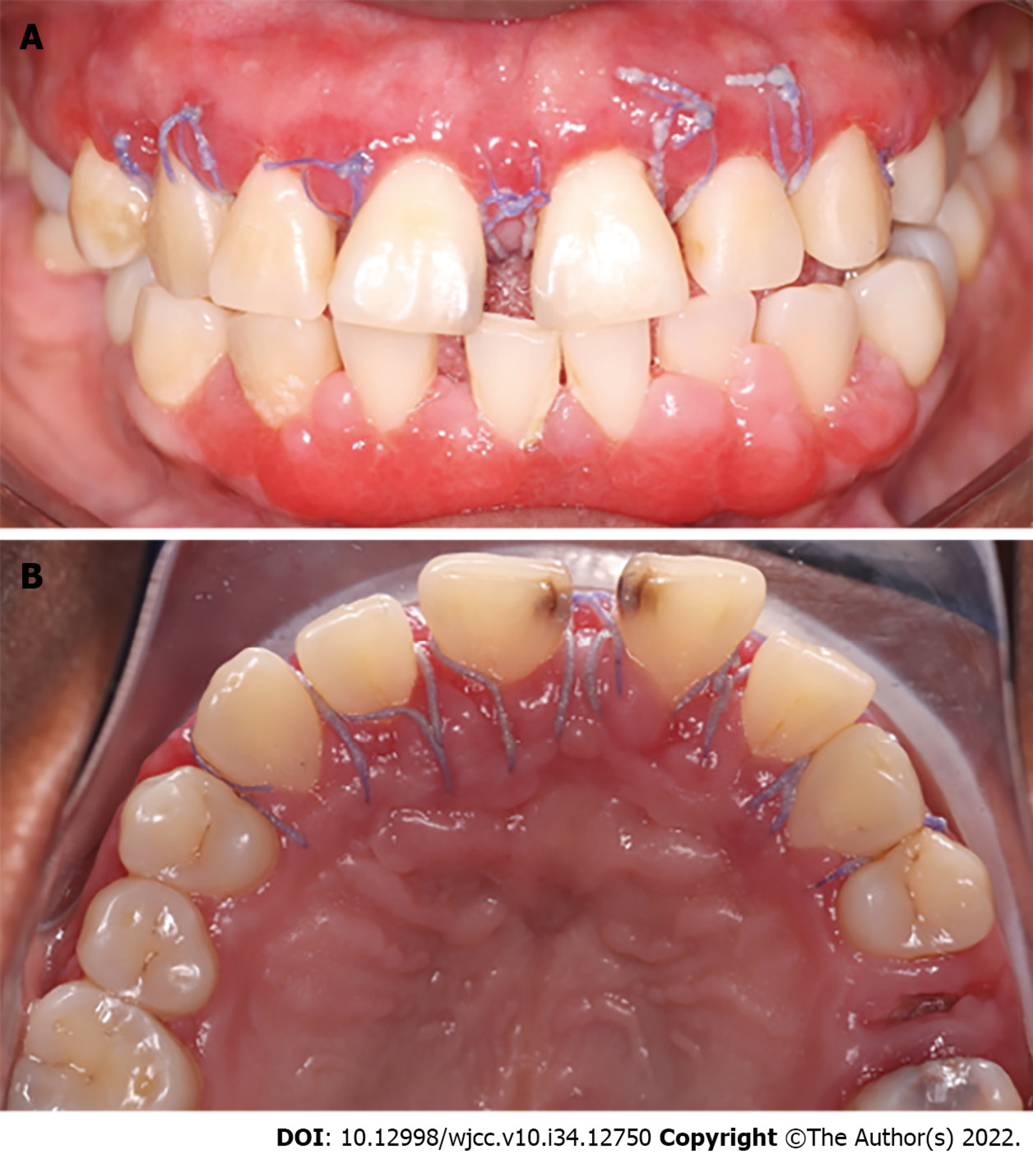
Figure 6 Corrective phase.
A: Vestibular reflected flap; B: Palatine reflected flap; C: Modified horizontal mattress sutures (vestibular view); D: Modified horizontal mattress sutures (palatal view).
Figure 7 Healing at postoperative 2 wk.
A slight inflammation was observed in line with the healing process, and the sutures were removed. A: Upper; B: Lower.
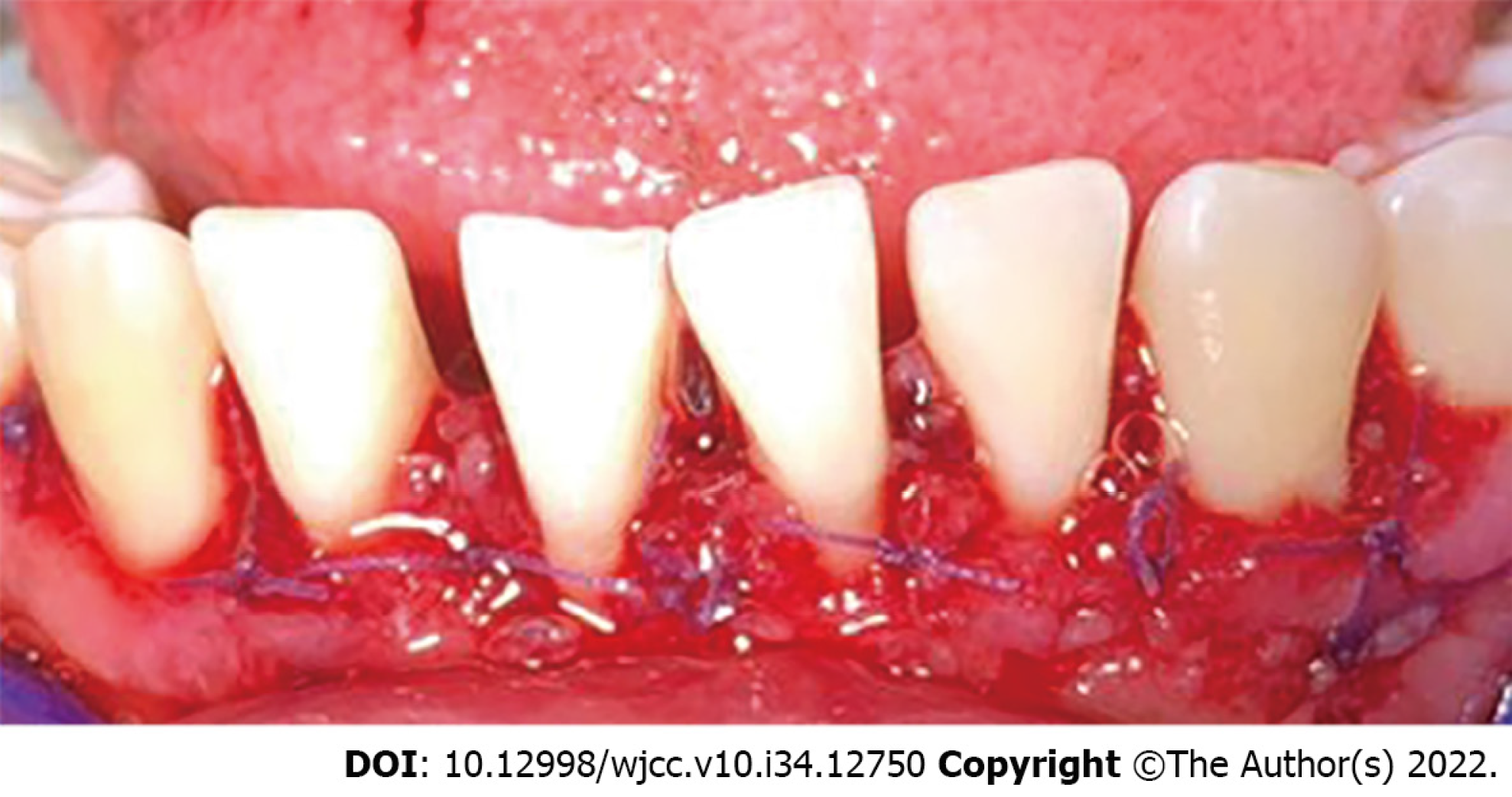
Figure 8 Postoperative view of the lower anterior sextant.
Depicting modified horizontal mattress sutures.
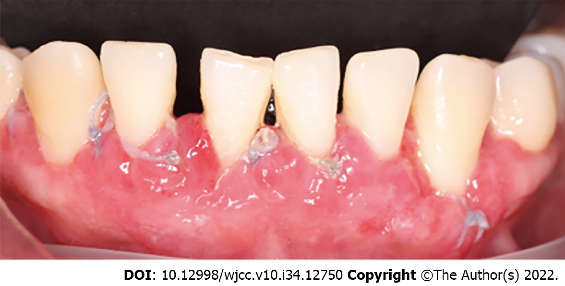
Figure 9 Lower anterior sextant at postoperative 2 wk.
Inflammation was observed in line with the healing process, and the sutures were removed.
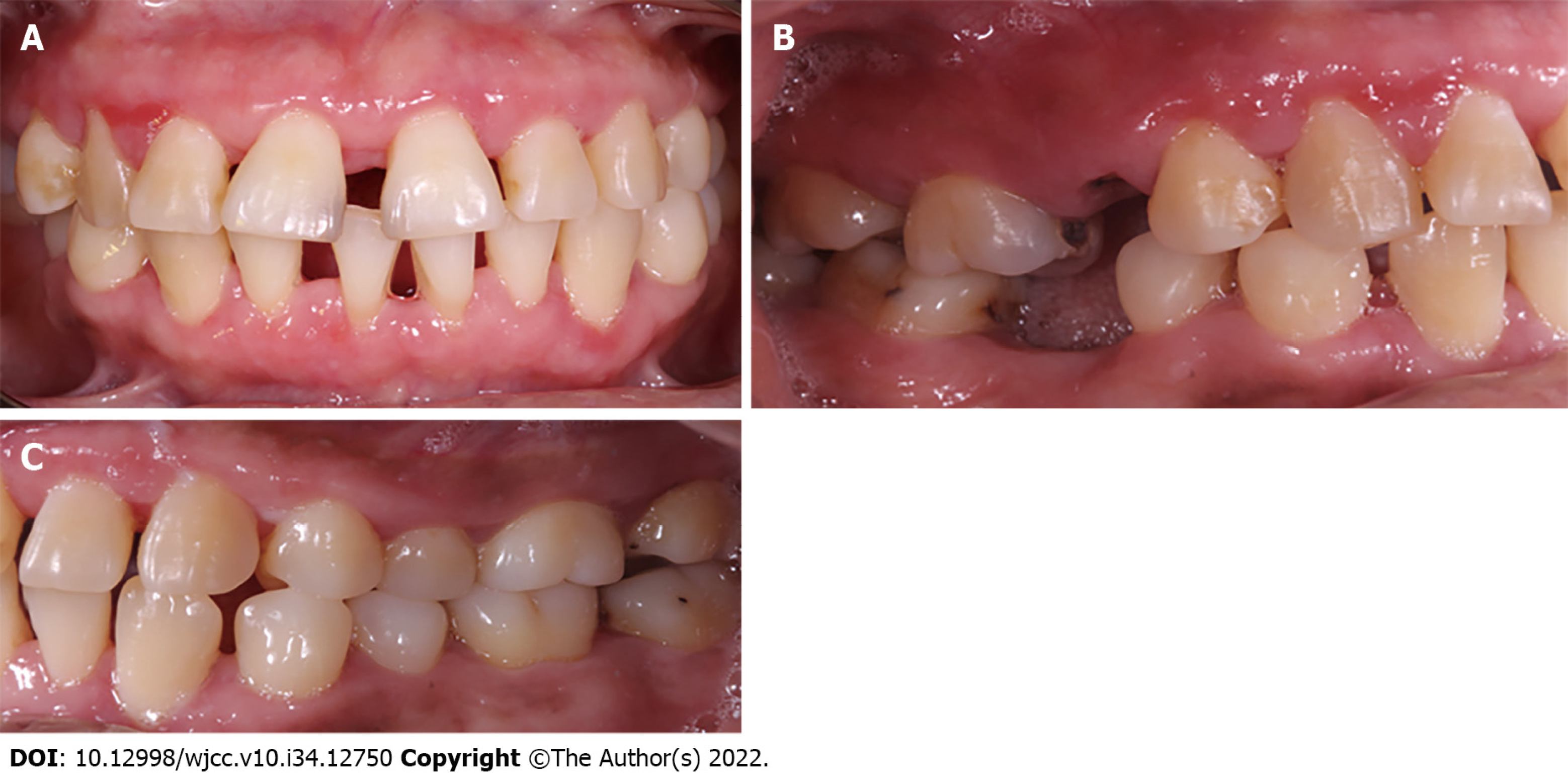
Figure 10 Photographs 3 mo after corrective phase.
The figure depicts a general improvement in tissue conditions. A: Inter-occlusal view; B: Right side view; C: Left side view.
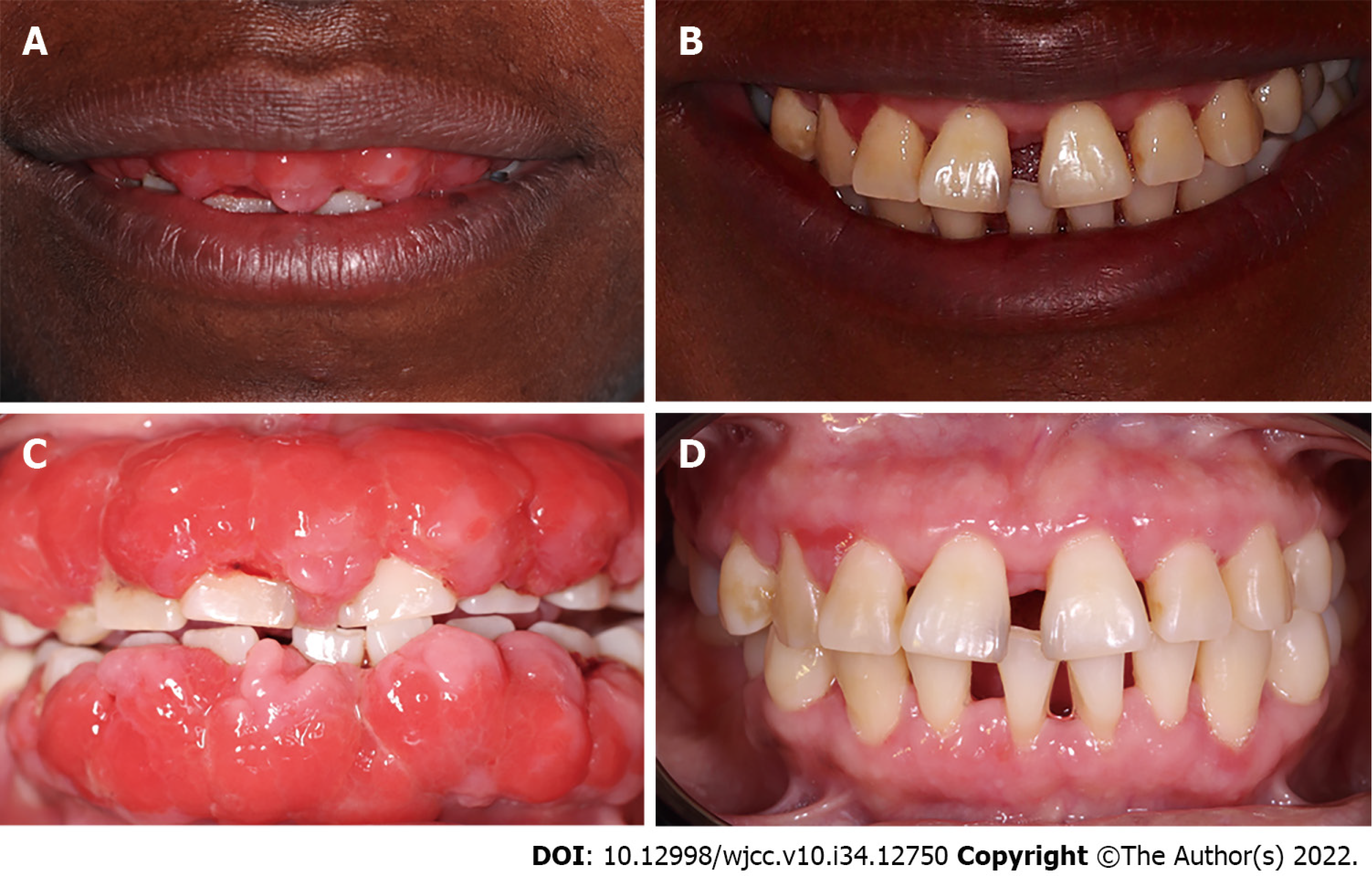
Figure 11 Comparative photographs.
A and C: Tissue conditions when the patient attended the periodontics clinic; B and D: Tissue status after three phases of periodontal treatment.
- Citation: Victory Rodríguez G, Ruiz Gutiérrez ADC, Gómez Sandoval JR, Lomelí Martínez SM. Gingival enlargement induced by cyclosporine in Medullary aplasia: A case report. World J Clin Cases 2022; 10(34): 12750-12760
- URL: https://www.wjgnet.com/2307-8960/full/v10/i34/12750.htm
- DOI: https://dx.doi.org/10.12998/wjcc.v10.i34.12750


