Copyright
©The Author(s) 2022.
World J Clin Cases. Nov 26, 2022; 10(33): 12395-12403
Published online Nov 26, 2022. doi: 10.12998/wjcc.v10.i33.12395
Published online Nov 26, 2022. doi: 10.12998/wjcc.v10.i33.12395
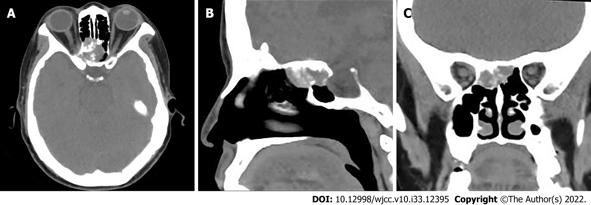
Figure 1 Computed tomography image of paranasal sinuses.
A: Cross-sectional images; B: Sagittal images; C: Coronal images.
Figure 2 Magnetic resonance imaging of the paranasal sinuses.
A: Heterogeneous low signal on T1-weighted images; B: Slightly high signal on fat-saturated T2-weighted images; C: Slightly low signal on D-weighted images.
Figure 3 Histopathological images.
A: Hematoxylin-eosin staining showed that diffuse distribution of undifferentiated round tumor cells, lamellar or focal arrangement, submucosal infiltrative growth, visible hemorrhage, necrosis, and invasion of bone (× 100); B-H: Immunohistochemical staining revealed positive nuclear protein in testis (B), AR1/AE3 (C), CK5/6 (D), CK8/18 (E), P40 (F), P63 (G), EGFR (H) (Envision × 200).
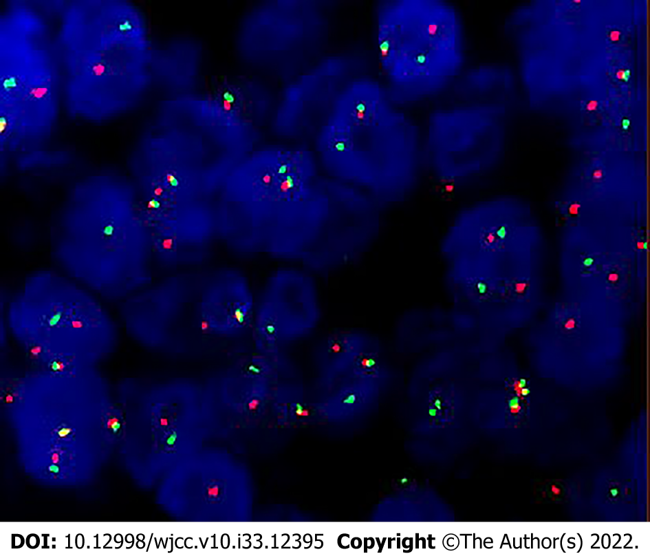
Figure 4 Nuclear protein in testis isolated probe fluorescence in situ hybridization.
Red and green signal separation in the tumor nucleus suggested a break in the gene.
Figure 5 Follow-up images of positron emission tomography/computed tomography and magnetic resonance imaging after paranasal sinuses nuclear protein in testis carcinoma resection.
A-D: Positron emission tomography/computed tomography (PET/CT) examination 1.5 mo after the surgery showed that the septal sinus, pterygoid sinus, right nasal cavity and anterior skull base soft tissue density shadows were radiolucent, maximum standardized uptake value was about 64.1, localized convexity into the orbits bilaterally, bilateral compression of the internal rectus muscle and optic nerve, and localized bone destruction in the septal sinus and pterygoid sinus (A, Maximum intensity projection image; B: Axial PET; C: Fused PET/CT; D: CT bone window images); E-H: 1.5 mo after surgery, magnetic resonance imaging (MRI) showed a recurrence of the tumor, measuring approximately 4.5 cm × 3.5 cm × 4.2 cm (E, T1-weighted image; F, T2-weighted image; G, D-weighted image; H, T1-weighted enhanced scan image); I-L: After 11 radiation treatments, the patient's MRI showed a significant tumor shrinkage, with a size of about 2.5 cm ×2.9 cm × 2.6 cm (I, T1-weighted image; J, T2-weighted image; K, D-weighted image; L, T1-weighted enhanced scan image); M-P: 6 mo after surgery, the follow-up evaluation revealed that the disease continued to progress, the tumor size was about 5.0 cm ×3.6 cm × 4.0 cm (M, T1-weighted image; N, T2-weighted image; O, D-weighted image; P, T1-weighted enhanced scan image).
- Citation: Huang WP, Gao G, Qiu YK, Yang Q, Song LL, Chen Z, Gao JB, Kang L. Multimodality imaging and treatment of paranasal sinuses nuclear protein in testis carcinoma: A case report. World J Clin Cases 2022; 10(33): 12395-12403
- URL: https://www.wjgnet.com/2307-8960/full/v10/i33/12395.htm
- DOI: https://dx.doi.org/10.12998/wjcc.v10.i33.12395


