Copyright
©The Author(s) 2022.
World J Clin Cases. Nov 16, 2022; 10(32): 12015-12021
Published online Nov 16, 2022. doi: 10.12998/wjcc.v10.i32.12015
Published online Nov 16, 2022. doi: 10.12998/wjcc.v10.i32.12015
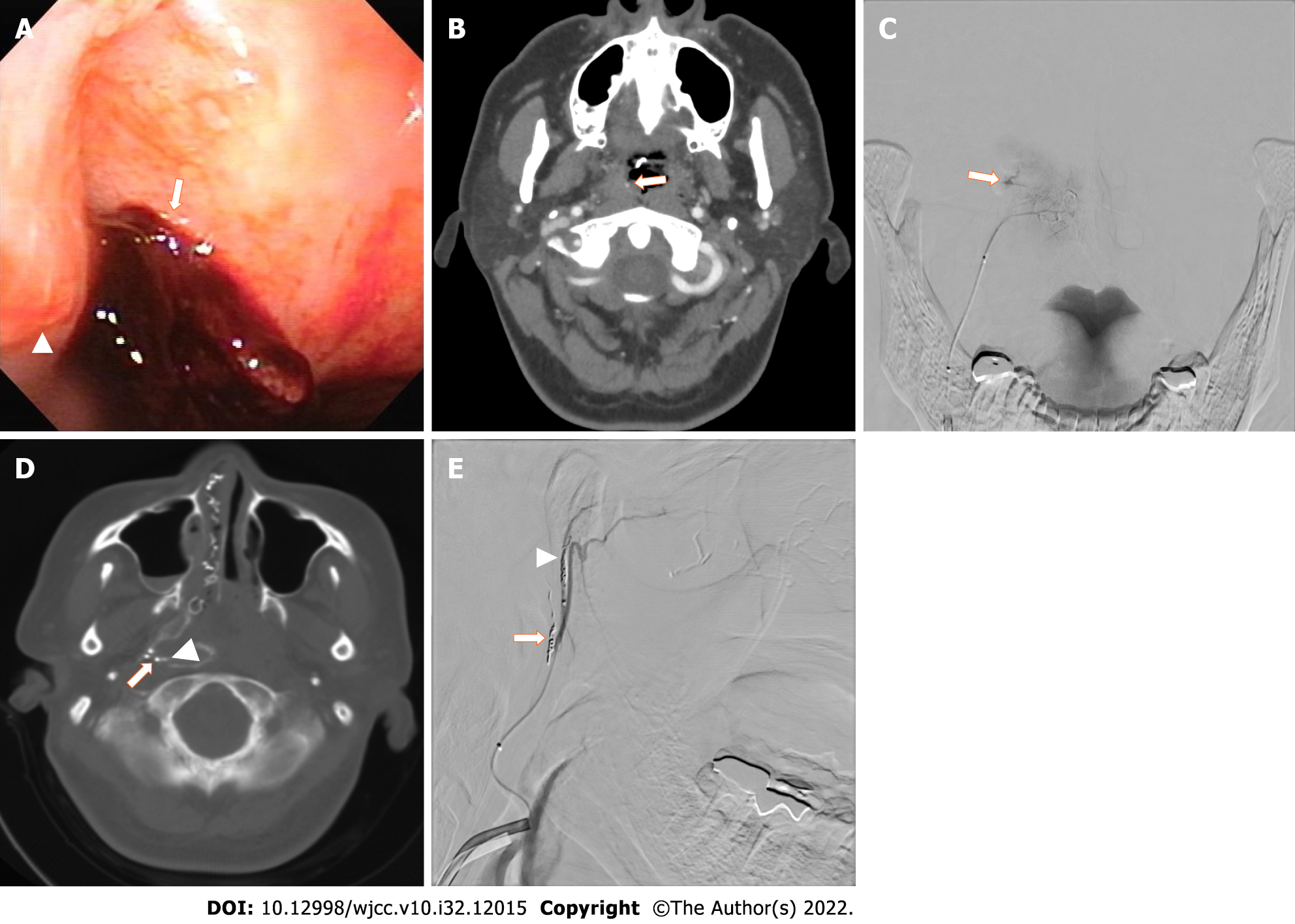
Figure 1 A 49-year-old Japanese woman who underwent transoral endoscopy under sedation for a medical check-up.
She suffered a hemorrhage from the nasopharynx due to the injury by the transnasal airway insertion. A: Trans-nasal endoscopy demonstrated formation of a clot at the right Rosenmuller fossa (arrow) posterior to the Torus tubarius (arrowhead); B: Contrast-enhanced computed tomography (CT) revealed extravasation in the nasopharynx (arrow); C: Angiography of the pharyngeal trunk of the ascending pharyngeal artery demonstrated extravasation (arrow); D: Xper CT imaging demonstrated an enhanced artery of the Rosenmuller fossa (arrow) and extravasation (arrowhead); E: Angiography demonstrated an embolized artery of the Rosenmuller fossa (arrow) and artery of Torus tubarius (arrowheads).
Figure 2 A 28-year-old Japanese woman who fell from the sixth floor of a building was transported to our hospital by ambulance.
A: Image of the patient during primary treatment in the emergency room. Due to excessive hemorrhage from the mouth, a transoral suction tube was placed in the mouth (arrow); B: Contrast-enhanced computed tomography demonstrated extravasation in the nasopharynx to hypopharynx (circles) and nasal cavity (arrows); C: Angiography of the pharyngeal trunk of the ascending pharyngeal artery demonstrated extravasation.
- Citation: Yunaiyama D, Takara Y, Kobayashi T, Muraki M, Tanaka T, Okubo M, Saguchi T, Nakai M, Saito K, Tsukahara K, Ishii Y, Homma H. Transcatheter arterial embolization for traumatic injury to the pharyngeal branch of the ascending pharyngeal artery: Two case reports. World J Clin Cases 2022; 10(32): 12015-12021
- URL: https://www.wjgnet.com/2307-8960/full/v10/i32/12015.htm
- DOI: https://dx.doi.org/10.12998/wjcc.v10.i32.12015


