Copyright
©The Author(s) 2022.
World J Clin Cases. Nov 16, 2022; 10(32): 12007-12014
Published online Nov 16, 2022. doi: 10.12998/wjcc.v10.i32.12007
Published online Nov 16, 2022. doi: 10.12998/wjcc.v10.i32.12007
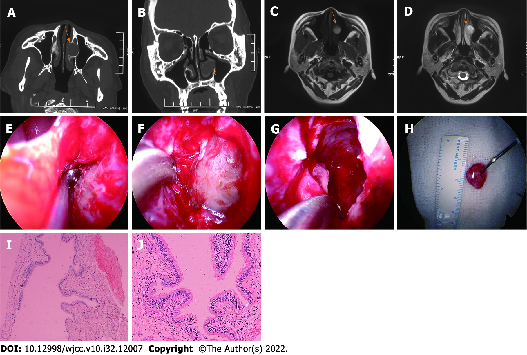
Figure 1 Case 1: A 55-year-old woman with an interior turbinate mucocele.
A: The inferior turbinate mucocele is shown on horizontal sinus computed tomography (CT); B: The inferior turbinate mucocele is shown on coronal sinus CT; C: The inferior turbinate mucocele is shown on T1 magnetic resonance imaging (MRI); D: The inferior turbinate mucocele is shown on T2 MRI; E: The bone wall of inferior turbinate mucocele is shown; F: The inferior turbinate mucocele lining is shown; G: The mucocele lining is completely removed; H: The inner layer diameter is about 1.2 cm; I: Hematoxylin and eosin (H&E) result of the mucocele lining is shown (× 40); J: H&E result of the mucocele lining is shown (× 100).
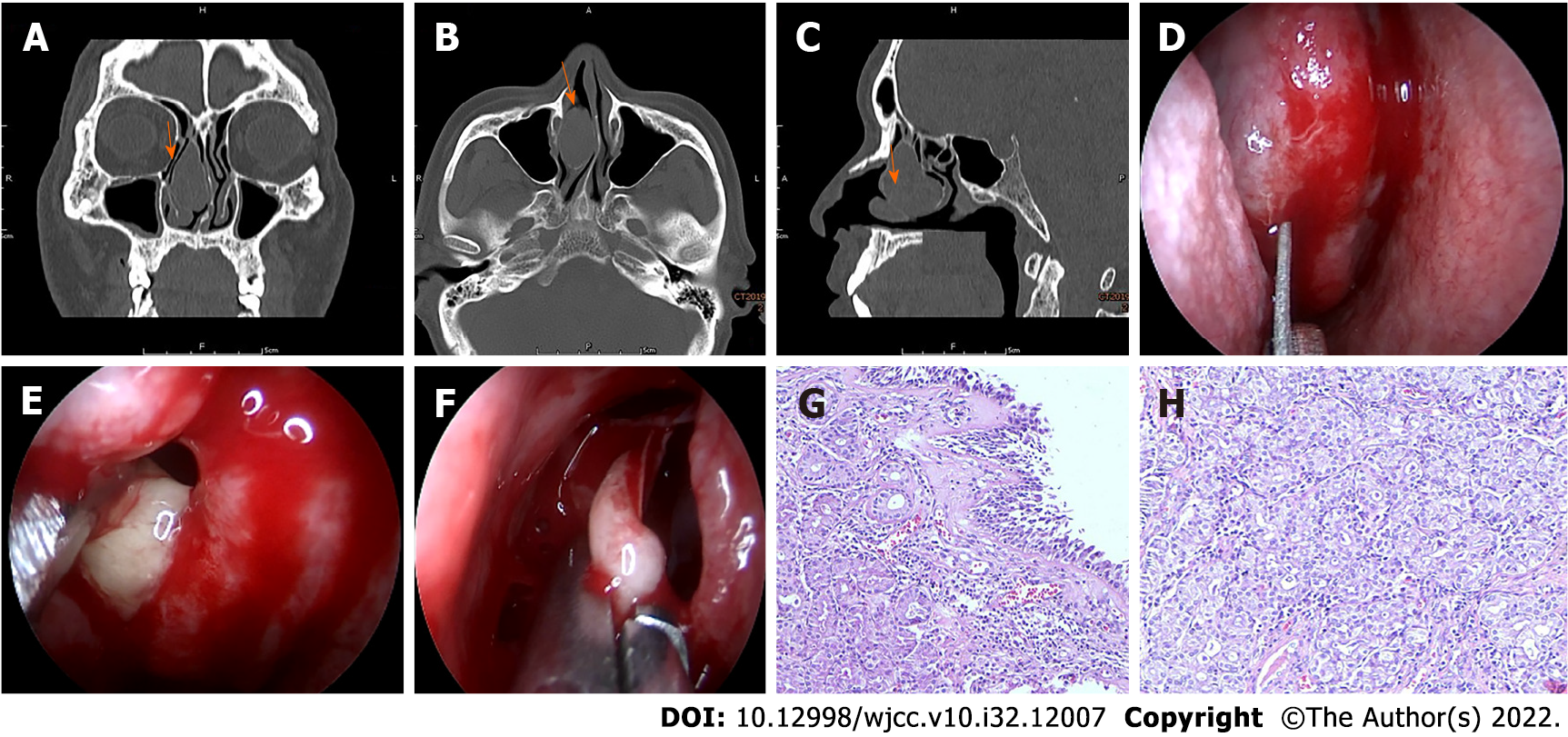
Figure 2 Case 2: A 58-year-old man with a middle turbinate pyogenic mucocele.
A: The middle turbinate pyogenic mucocele is shown on coronal sinus computed tomography (CT); B: The middle turbinate pyogenic mucocele is shown on horizontal sinus CT; C: The middle turbinate pyogenic mucocele is shown on sagittal sinus CT; D: The full-thickness longitudinal incision was made at the front end of the right middle turbinate; E: The white pus oozing out from middle turbinate pyogenic mucocele; F: The medial lamellae of the pyogenic mucocele are resected; G and H: Hematoxylin and eosin result of the medial lamellae of the pyogenic mucocele is shown (× 40).
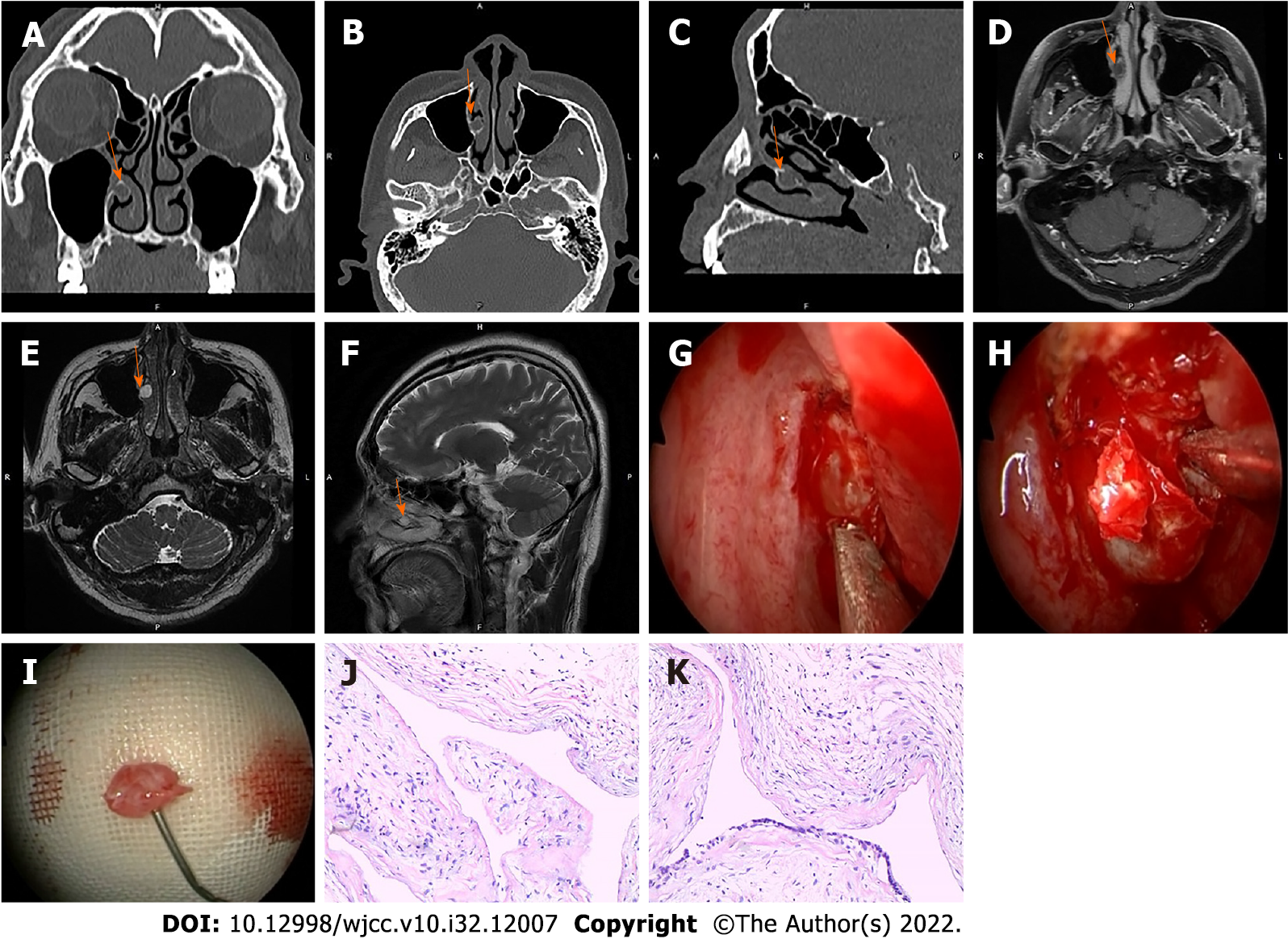
Figure 3 Case 3: A 50-year-old man with an inferior turbinate mucocele.
A: The inferior turbinate mucocele is shown on coronal sinus computed tomography (CT); B: The inferior turbinate mucocele is shown on horizontal sinus CT; C: The inferior turbinate mucocele is shown on sagittal sinus CT; D: The inferior turbinate mucocele is shown on T2 horizontal magnetic resonance imaging (MRI); E: The inferior turbinate mucocele is shown on T1 horizontal MRI; F: The inferior turbinate mucocele is shown on T2 coronal MRI; G: The bone wall of inferior turbinate mucocele is shown; H: The mucocele lining is completely removed; I: The inner layer diameter is about 0.8 cm; J and K: Hematoxylin and eosin result of the mucocele lining is shown (× 40).
- Citation: Sun SJ, Chen AP, Wan YZ, Ji HZ. Endoscopic nasal surgery for mucocele and pyogenic mucocele of turbinate: Three case reports. World J Clin Cases 2022; 10(32): 12007-12014
- URL: https://www.wjgnet.com/2307-8960/full/v10/i32/12007.htm
- DOI: https://dx.doi.org/10.12998/wjcc.v10.i32.12007


