Copyright
©The Author(s) 2022.
World J Clin Cases. Nov 16, 2022; 10(32): 12000-12006
Published online Nov 16, 2022. doi: 10.12998/wjcc.v10.i32.12000
Published online Nov 16, 2022. doi: 10.12998/wjcc.v10.i32.12000
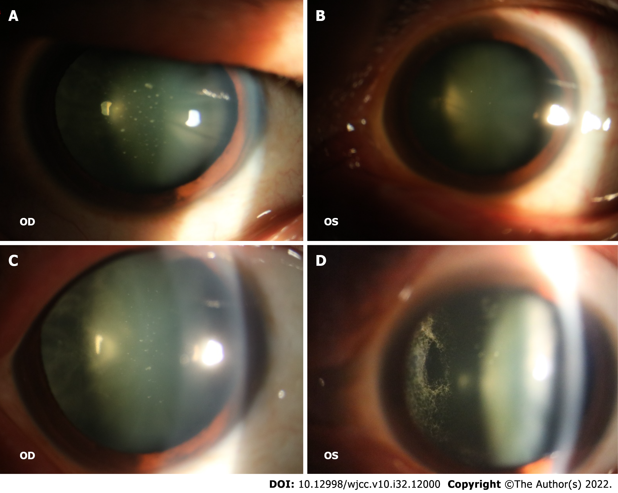
Figure 1 Physical examination.
A and B: White spots on the posterior surface of the lens in both eyes, which were similar to foot plates; C and D: Fundus examination revealed vitreous opacity in both eyes, with a noticeable spider-web-like white cord.
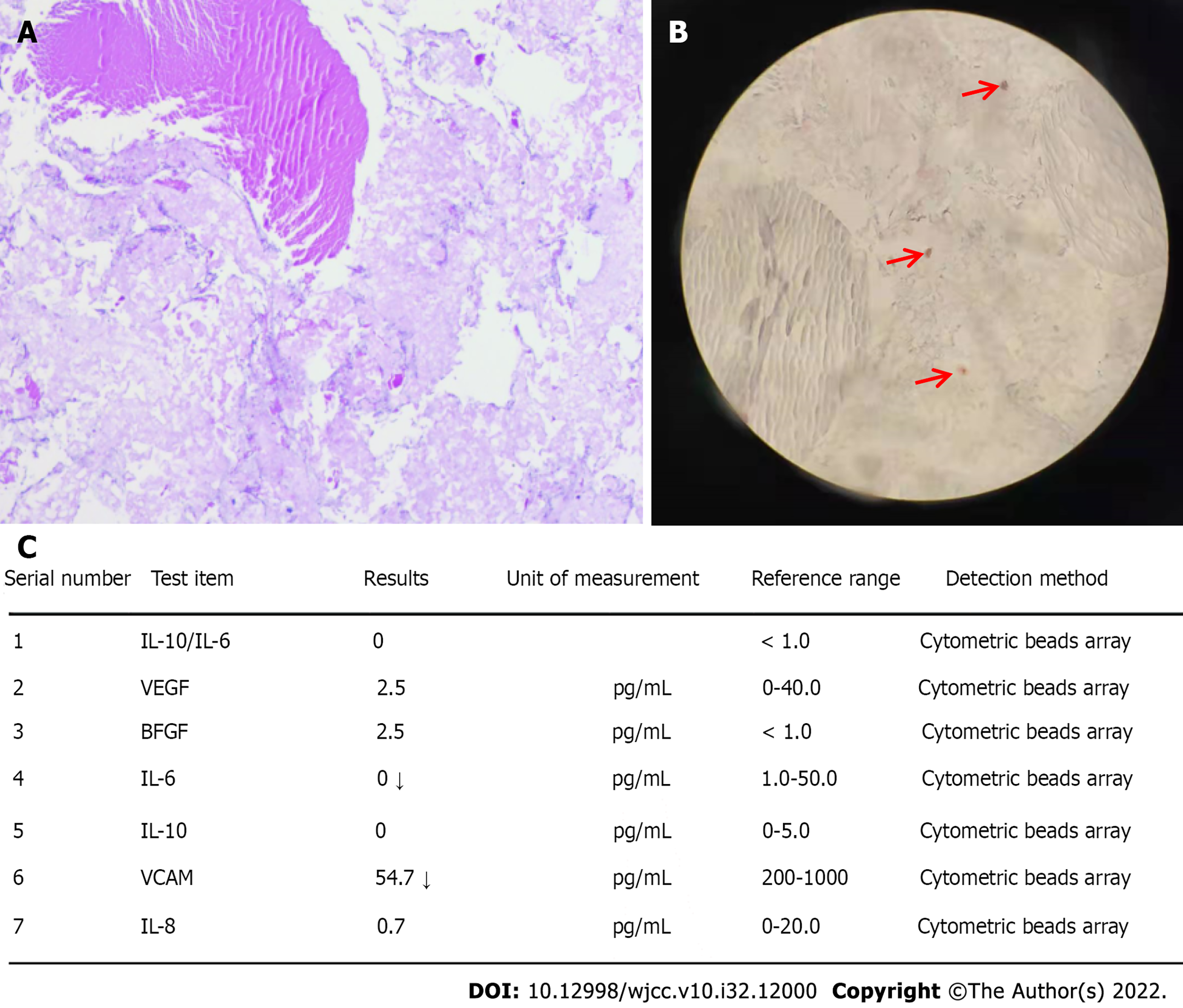
Figure 2 Laboratory examinations.
A: Hematoxylin and eosin staining revealed a waxy light eosinophil; B: Congo red staining was positive (at the arrow); C: Interleukin (IL)-6 and IL-10 levels were normal.
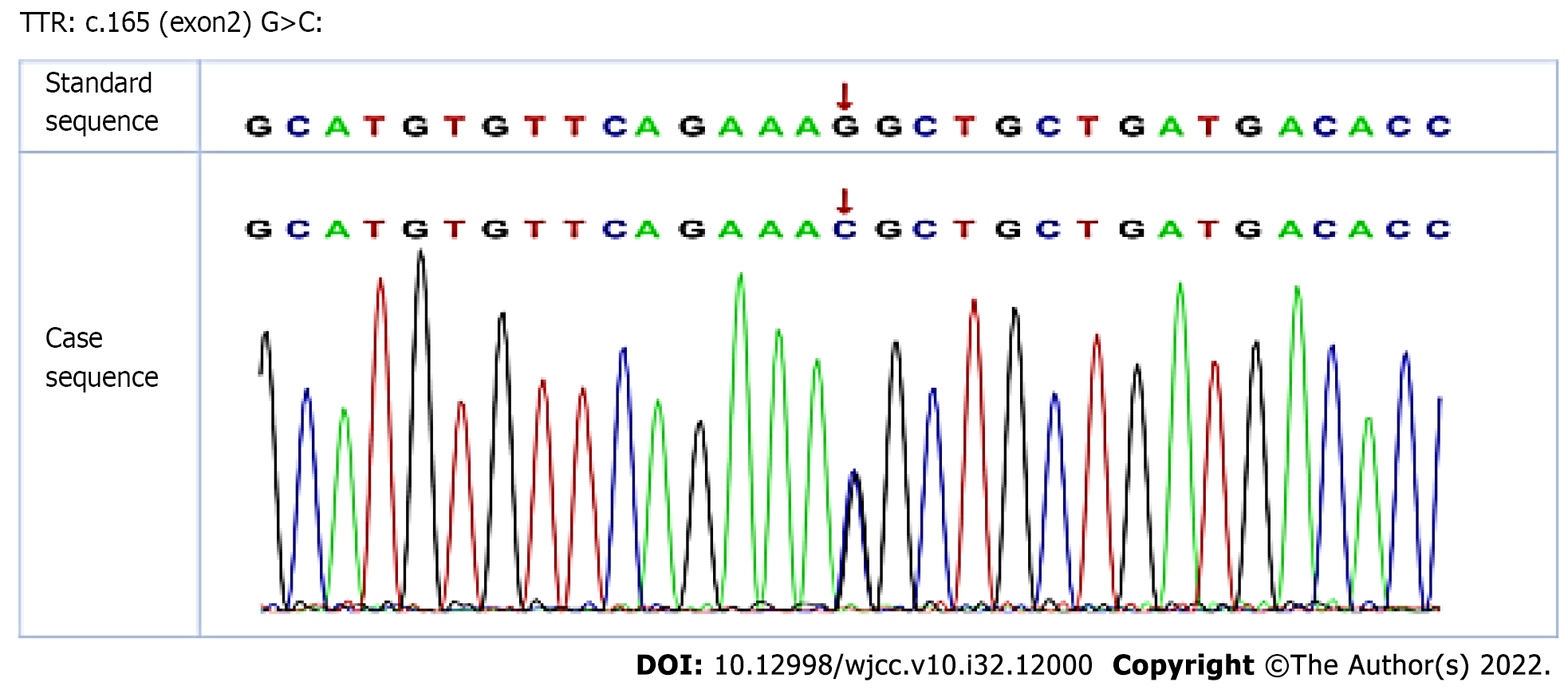
Figure 3 A chromatogram revealing the genetic sequencing result of the patient.
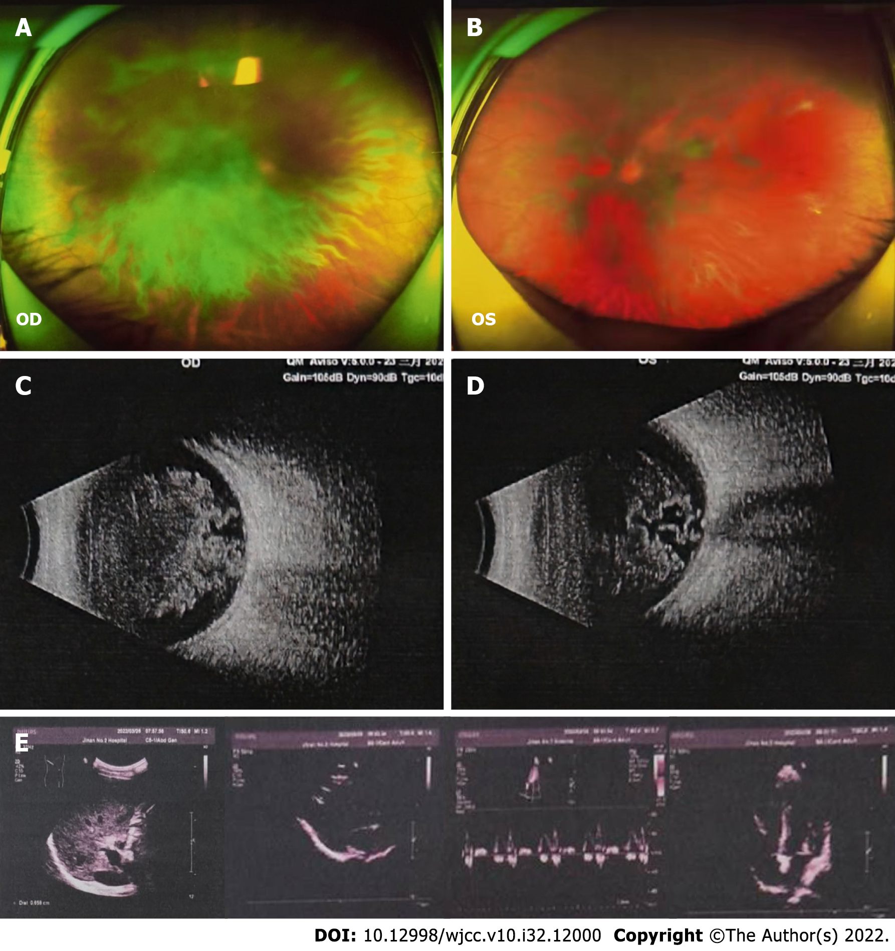
Figure 4 Imaging examinations.
A-E: Fundus color photography (A and B) and ultrasound examination (C and D) showed obvious opacity in bilateral vitreous bodies. There was no obvious abnormality of the heart or abdomen by color ultrasound (E).
- Citation: Tan Y, Tao Y, Sheng YJ, Zhang CM. Vitreous amyloidosis caused by a Lys55Asn variant in transthyretin: A case report. World J Clin Cases 2022; 10(32): 12000-12006
- URL: https://www.wjgnet.com/2307-8960/full/v10/i32/12000.htm
- DOI: https://dx.doi.org/10.12998/wjcc.v10.i32.12000


