Copyright
©The Author(s) 2022.
World J Clin Cases. Nov 16, 2022; 10(32): 11974-11979
Published online Nov 16, 2022. doi: 10.12998/wjcc.v10.i32.11974
Published online Nov 16, 2022. doi: 10.12998/wjcc.v10.i32.11974
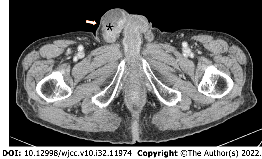
Figure 1 Computed tomography urography image showing a mixed solid-cystic mass in the right testicular and epididymal area.
Figure 2 Histological analysis of testicular and epididymal tumor samples.
A and B: Hematoxylin and eosin staining revealed poorly differentiated adenocarcinoma in the testis and epididymis (magnification: × 400); C and D: Immunohistochemistry revealed that the tumor cells were positive for pancytokeratins and caudal related homeodomain transcription 2 (magnification: × 200).
- Citation: Ji JJ, Guan FJ, Yao Y, Sun LJ, Zhang GM. Testis and epididymis–unusual sites of metastatic gastric cancer: A case report and review of the literature. World J Clin Cases 2022; 10(32): 11974-11979
- URL: https://www.wjgnet.com/2307-8960/full/v10/i32/11974.htm
- DOI: https://dx.doi.org/10.12998/wjcc.v10.i32.11974


