Copyright
©The Author(s) 2022.
World J Clin Cases. Nov 16, 2022; 10(32): 11967-11973
Published online Nov 16, 2022. doi: 10.12998/wjcc.v10.i32.11967
Published online Nov 16, 2022. doi: 10.12998/wjcc.v10.i32.11967
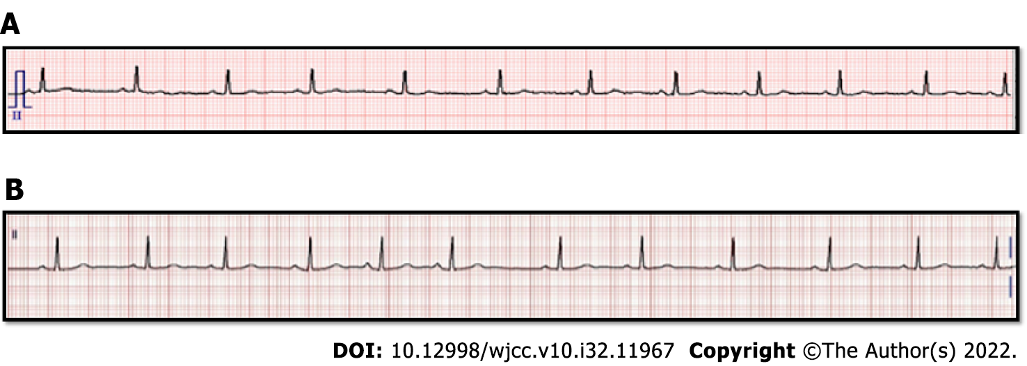
Figure 1 Periopreative electrocardiogram of the patient.
A: Preoperative electrocardiogram (ECG), Sinus rhythm, heart rate (HR) 68bpm, QT/QTc 361/384 ms; B: Postoperative ECG, showing the premature atrial complexes, Sinus rhythm with premature atrial complexes HR 71bpm, QT/QTc 392/425 ms.
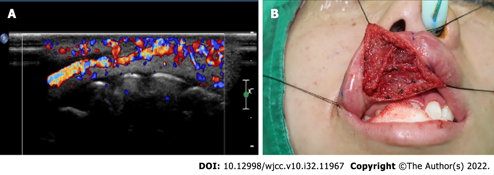
Figure 2 Images of the patient.
A: Ultrasound image: Increased vascularity in the right upper lip, probably vascular malformation; B: Intraoperative picture: Tracing with black silk to expose the surgical site during the approach to the surgical site.
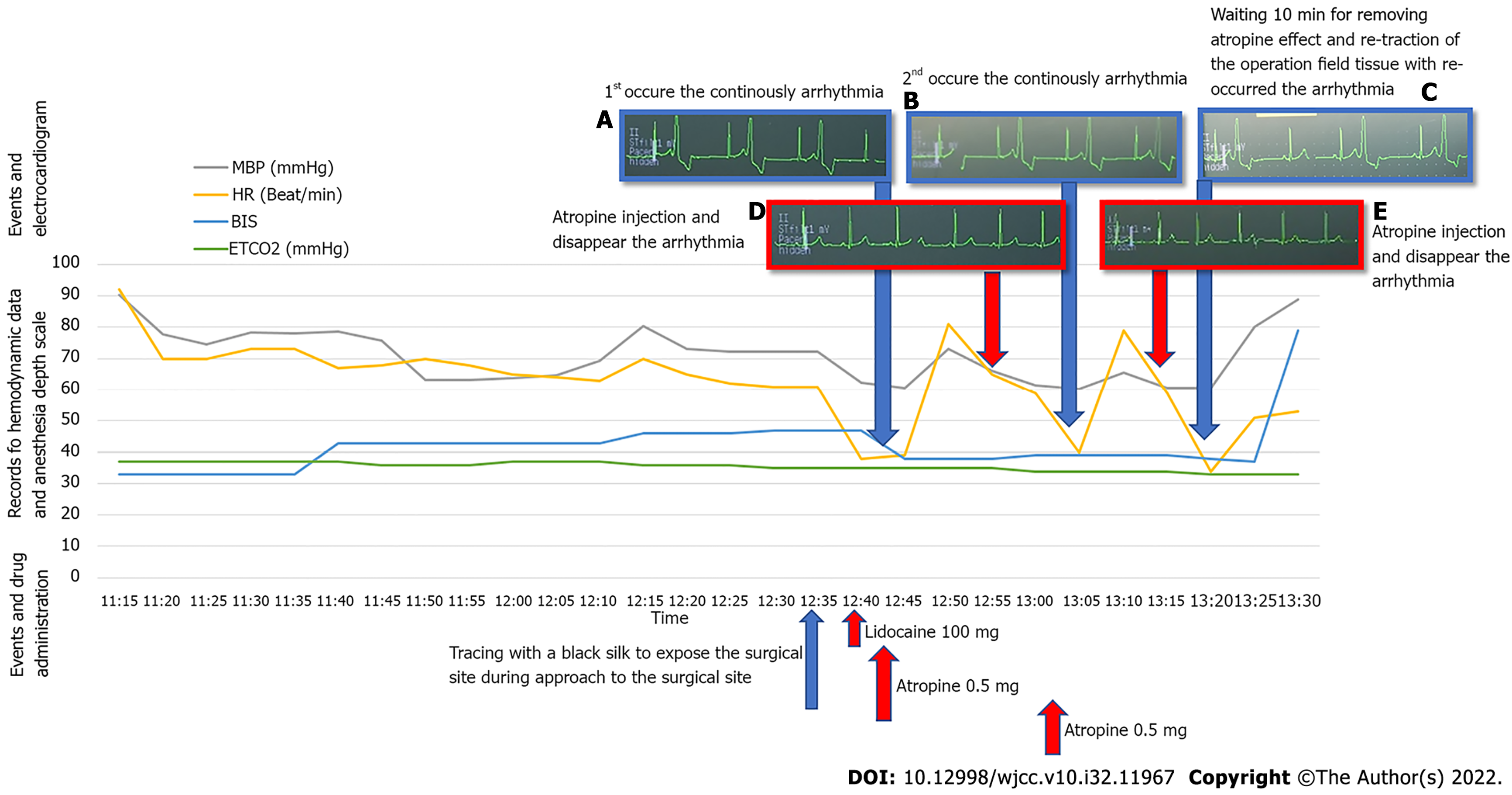
Figure 3 Hemodynamic data and events with drug administration and electrocardiogram in chronological order.
A: First, arrythmia occurred continuously; B: Atropine injection was administered, and arrhythmia with bradycardia disappeared; C: Second arrhythmia occurred continuously; D: Atropine injection was administered, and arrhythmia with bradycardia disappeared; E: Wait for 10 min for the atropine to take effect and arrhythmia reoccurred with re-traction of the surgical field tissue. MBP: Mean blood pressure; HR: Heart rate; BP: Blood pressure; BIS: Bispectral index; ETCO2: End-tidal CO2.
- Citation: Cho SY, Jang BH, Jeon HJ, Kim DJ. Repeated ventricular bigeminy by trigeminocardiac reflex despite atropine administration during superficial upper lip surgery: A case report. World J Clin Cases 2022; 10(32): 11967-11973
- URL: https://www.wjgnet.com/2307-8960/full/v10/i32/11967.htm
- DOI: https://dx.doi.org/10.12998/wjcc.v10.i32.11967


