Copyright
©The Author(s) 2022.
World J Clin Cases. Nov 16, 2022; 10(32): 11949-11954
Published online Nov 16, 2022. doi: 10.12998/wjcc.v10.i32.11949
Published online Nov 16, 2022. doi: 10.12998/wjcc.v10.i32.11949
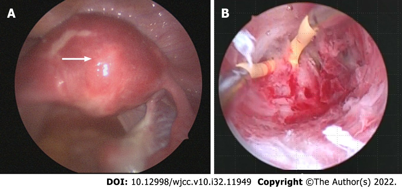
Figure 1 Intraoperative findings.
A: The hysteroscope light source was kept visible during laparoscopic observation; B: Blunt dissection of adhesion tissue was performed using a monopolar electric scalpel.
Figure 2
Postoperative course.
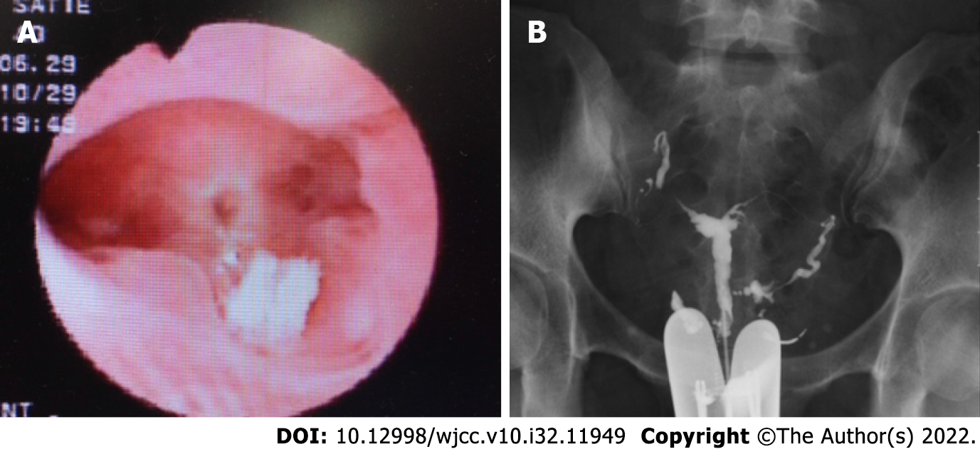
Figure 3 Postoperative findings.
A: Hysteroscopy shows no intrauterine adhesions; endometrial regeneration is evident; B: Hysterosalpingography demonstrates contrast medium reaching the fimbriae of both fallopian tubes from the uterine cavity and leaking into the abdominal cavity.
- Citation: Kakinuma T, Kakinuma K, Matsuda Y, Ohwada M, Yanagida K. Successful live birth following hysteroscopic adhesiolysis under laparoscopic observation for Asherman’s syndrome: A case report. World J Clin Cases 2022; 10(32): 11949-11954
- URL: https://www.wjgnet.com/2307-8960/full/v10/i32/11949.htm
- DOI: https://dx.doi.org/10.12998/wjcc.v10.i32.11949


