Copyright
©The Author(s) 2022.
World J Clin Cases. Nov 16, 2022; 10(32): 11898-11907
Published online Nov 16, 2022. doi: 10.12998/wjcc.v10.i32.11898
Published online Nov 16, 2022. doi: 10.12998/wjcc.v10.i32.11898
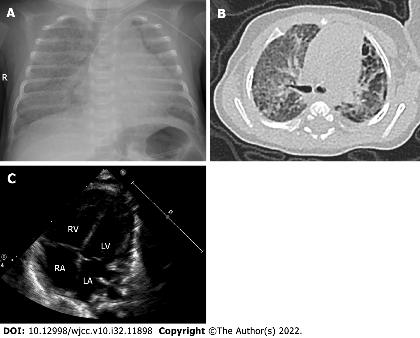
Figure 1 The imaging characteristics of the infant at 65 d after birth.
A: Chest X-ray of the infant. Blurred lung texture, broad reduction in light transmission in both lungs, extensive lamellar dense shadow, and blurred mesh shadow were seen in both lungs, which suggested interstitial changes in both lungs; B: Computed tomography scan of the infant. Uneven transmission of both lungs with small capsule cavity locally, grind glass, dots, and patchy shadows scattered in both lungs with latticed changes locally. The interstitial abnormality in both lungs suggests bronchopulmonary dysplasia; C: Echocardiogram of the infant. Enlargement of the right ventricle and atrium was seen as well as thickening of the right ventricle anterior wall. The estimated pulmonary artery pressure was 47 mmHg according to the tricuspid regurgitation.
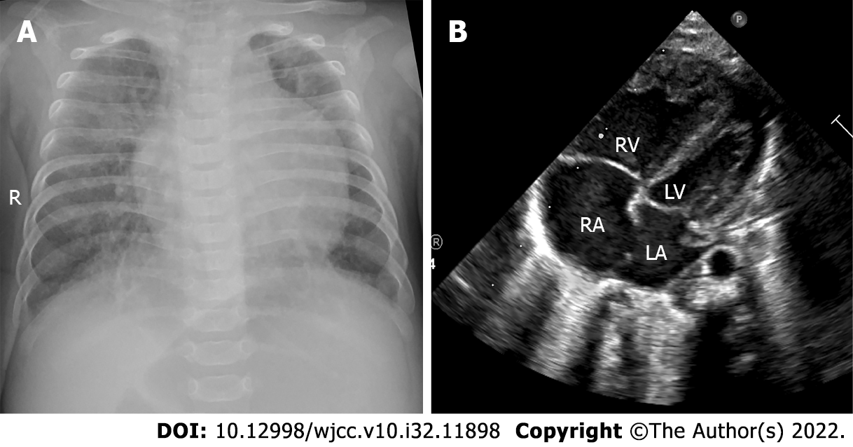
Figure 2 The imaging characteristics of the infant at 151 d after birth.
A: Chest X-ray of the infant. Uneven transmission of both lungs, blurred lung texture, grind glass, latticed and patchy shadows scattered in both lungs, increased cardiac shadow, and a heart to chest ratio of 0.63 suggest inflammation of both lungs with interstitial changes; B: Echocardiogram of the infant. Right heart enlarged, left heat narrowed. The estimated pulmonary artery pressure was 88 mmHg according to the tricuspid regurgitation, suggesting severe pulmonary hypertension.
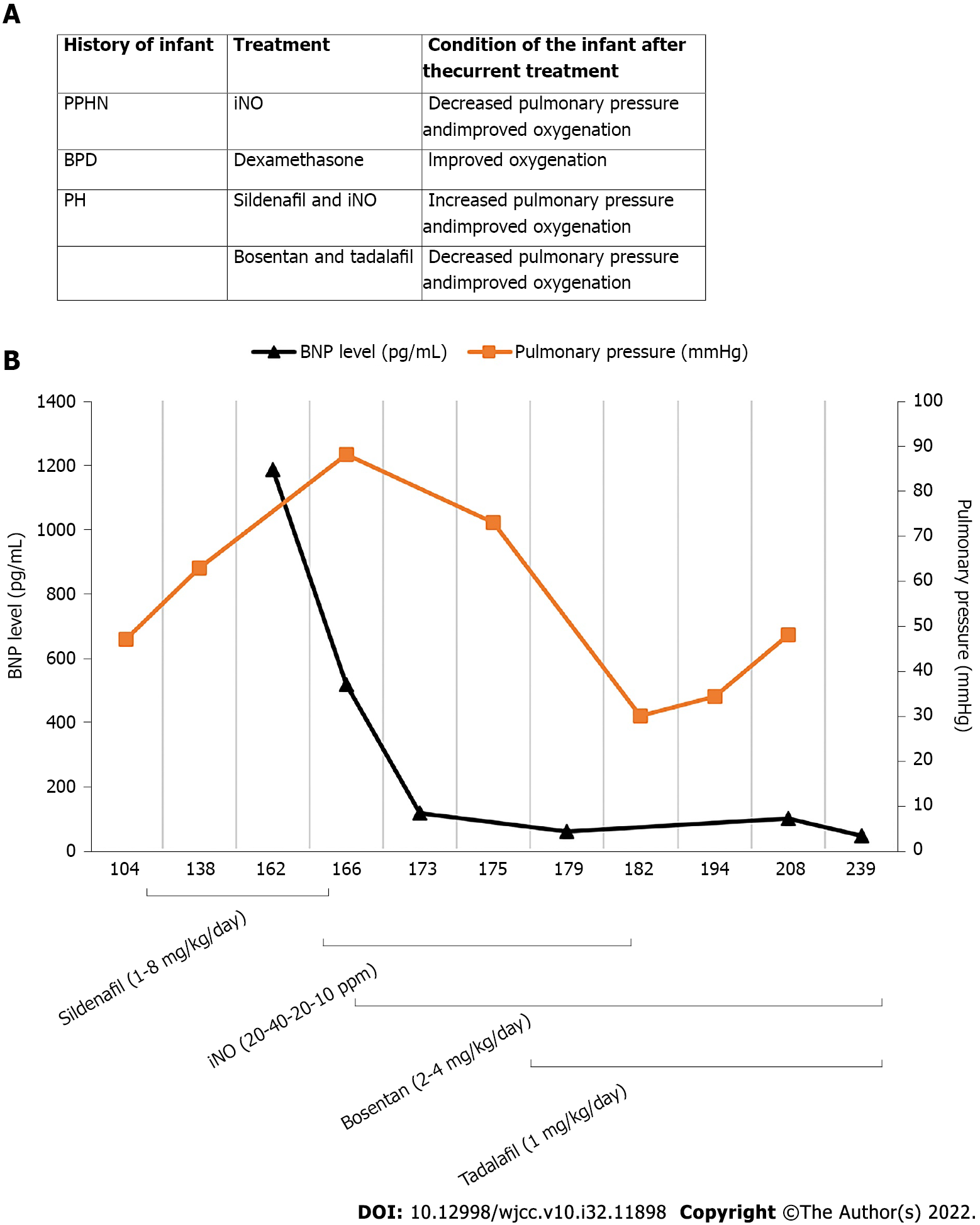
Figure 3 Clinical course of the patient.
A: Brief treatment chart of the infant; B: Serial changes in the B-type natriuretic peptide level and pulmonary arterial pressure, and the clinical course of pulmonary hypertension (PH) of the patient. BPD: Bronchopulmonary dysplasia; iNO: Inhaled nitric oxide; PPHN: Persistent PH of the newborn.
- Citation: Li J, Zhao J, Yang XY, Shi J, Liu HT. Successful treatment of pulmonary hypertension in a neonate with bronchopulmonary dysplasia: A case report and literature review. World J Clin Cases 2022; 10(32): 11898-11907
- URL: https://www.wjgnet.com/2307-8960/full/v10/i32/11898.htm
- DOI: https://dx.doi.org/10.12998/wjcc.v10.i32.11898


