Copyright
©The Author(s) 2022.
World J Clin Cases. Nov 16, 2022; 10(32): 11845-11852
Published online Nov 16, 2022. doi: 10.12998/wjcc.v10.i32.11845
Published online Nov 16, 2022. doi: 10.12998/wjcc.v10.i32.11845
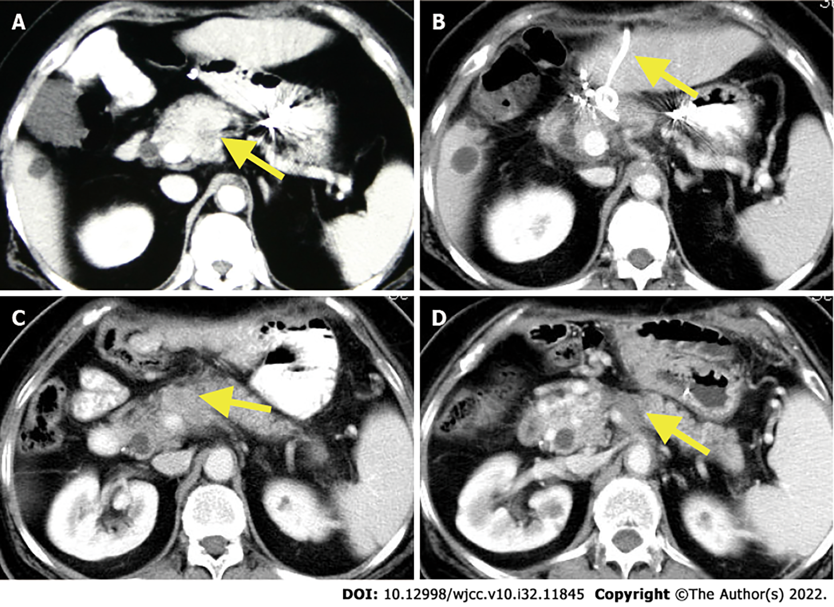
Figure 1 Contrast-enhanced computed tomography of the tumor lesions.
A: Primary tumor (arrow) lesion before the initial diagnosis; B: 1-mo follow-up contrast-enhanced computed tomography (CECT): The patient had a pancreatic fistula after radiofrequency ablation (RFA) and the percutaneous transhepatic drainage tube is shown in the figure (arrow); C: 8-mo follow-up CECT: Local recurrence was found (arrow); D: 8-year follow-up CECT after repeated RFA: A necrotic area that was suggestive of an ablated tumor (arrow).
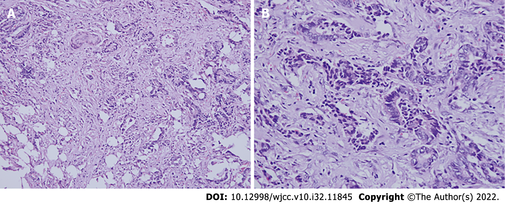
Figure 2 Patient’s tumor, hematoxylin and eosin staining.
A: Invasive mucinous adenocarcinoma with interstitial invasion (200 ×); B: Poorly differentiated pancreatic ductal adenocarcinoma (400 ×). Most of the glands were arranged like ducts and some glands were arranged like cords.
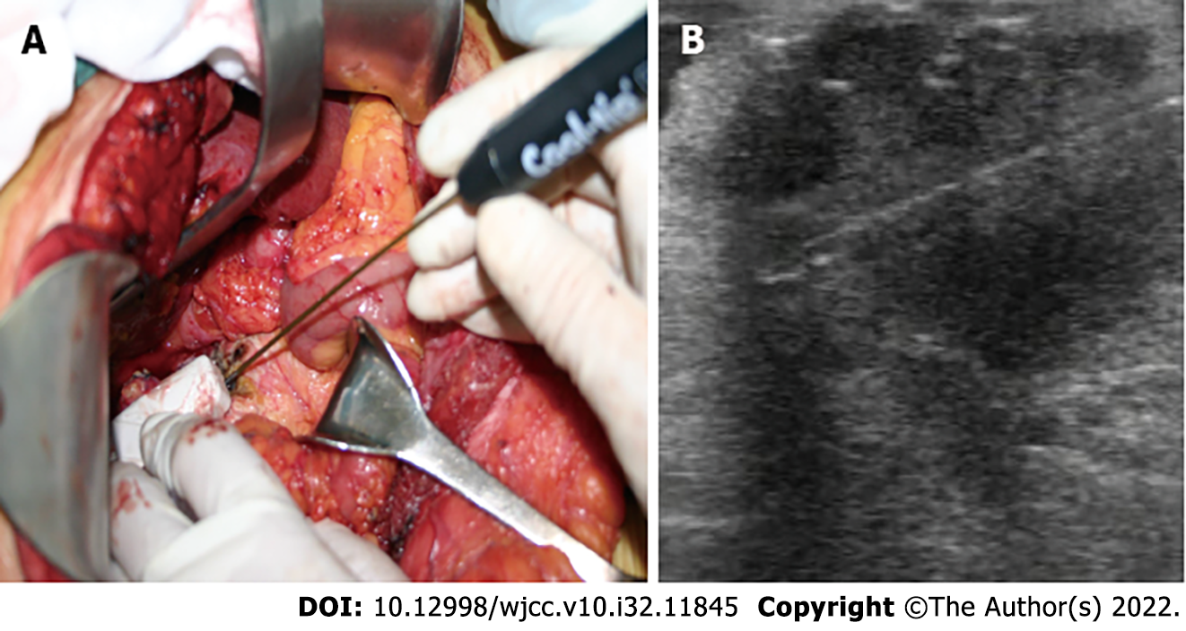
Figure 3 Operative findings of the first radiofrequency ablation.
A: Intraoperative placement of radiofrequency ablation (RFA) probe under direct ultrasound guidance; B: The tip for RFA was placed inside the tumor under intraoperative ultrasound guidance.
- Citation: Zhang JY, Ding JM, Zhou Y, Jing X. Nine-year survival of a 60-year-old woman with locally advanced pancreatic cancer under repeated open approach radiofrequency ablation: A case report. World J Clin Cases 2022; 10(32): 11845-11852
- URL: https://www.wjgnet.com/2307-8960/full/v10/i32/11845.htm
- DOI: https://dx.doi.org/10.12998/wjcc.v10.i32.11845


