Copyright
©The Author(s) 2022.
World J Clin Cases. Oct 26, 2022; 10(30): 11178-11184
Published online Oct 26, 2022. doi: 10.12998/wjcc.v10.i30.11178
Published online Oct 26, 2022. doi: 10.12998/wjcc.v10.i30.11178
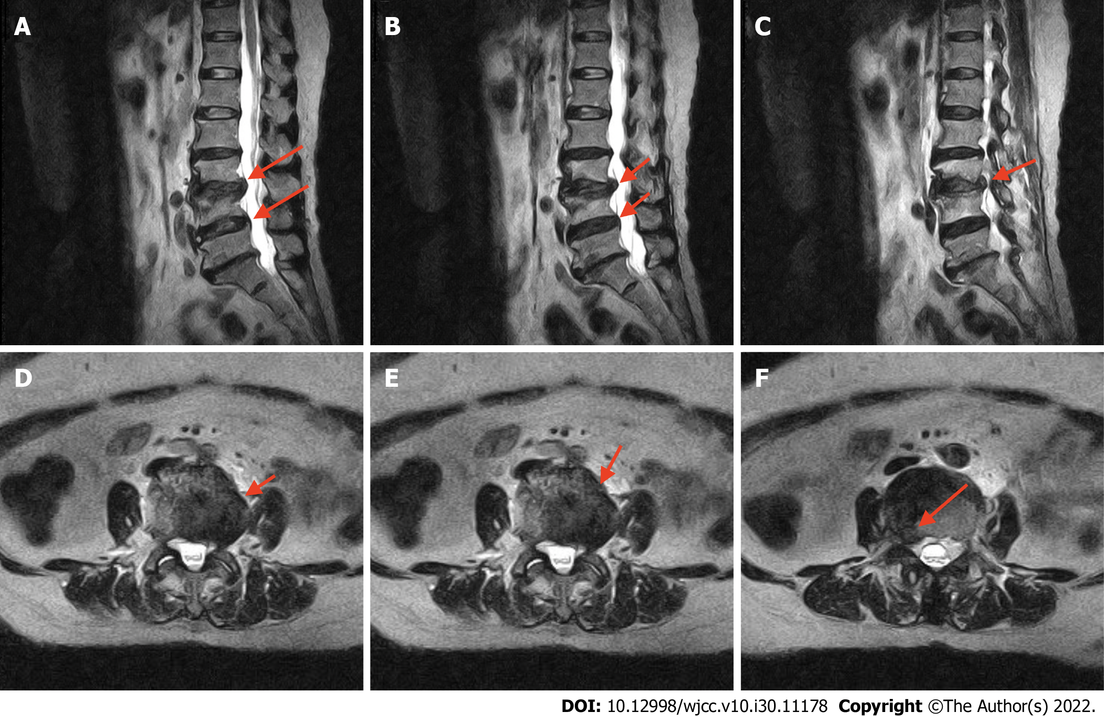
Figure 1 Preoperative T2-weighted magnetic resonance imaging.
The lateral and sagittal views of the collapsed disc height at the L2-3 and L3-4 Levels and a bulging disk compressing the thecal sac and right neural foramen are visible. A-C: Lateral views; D-F: Sagittal views.
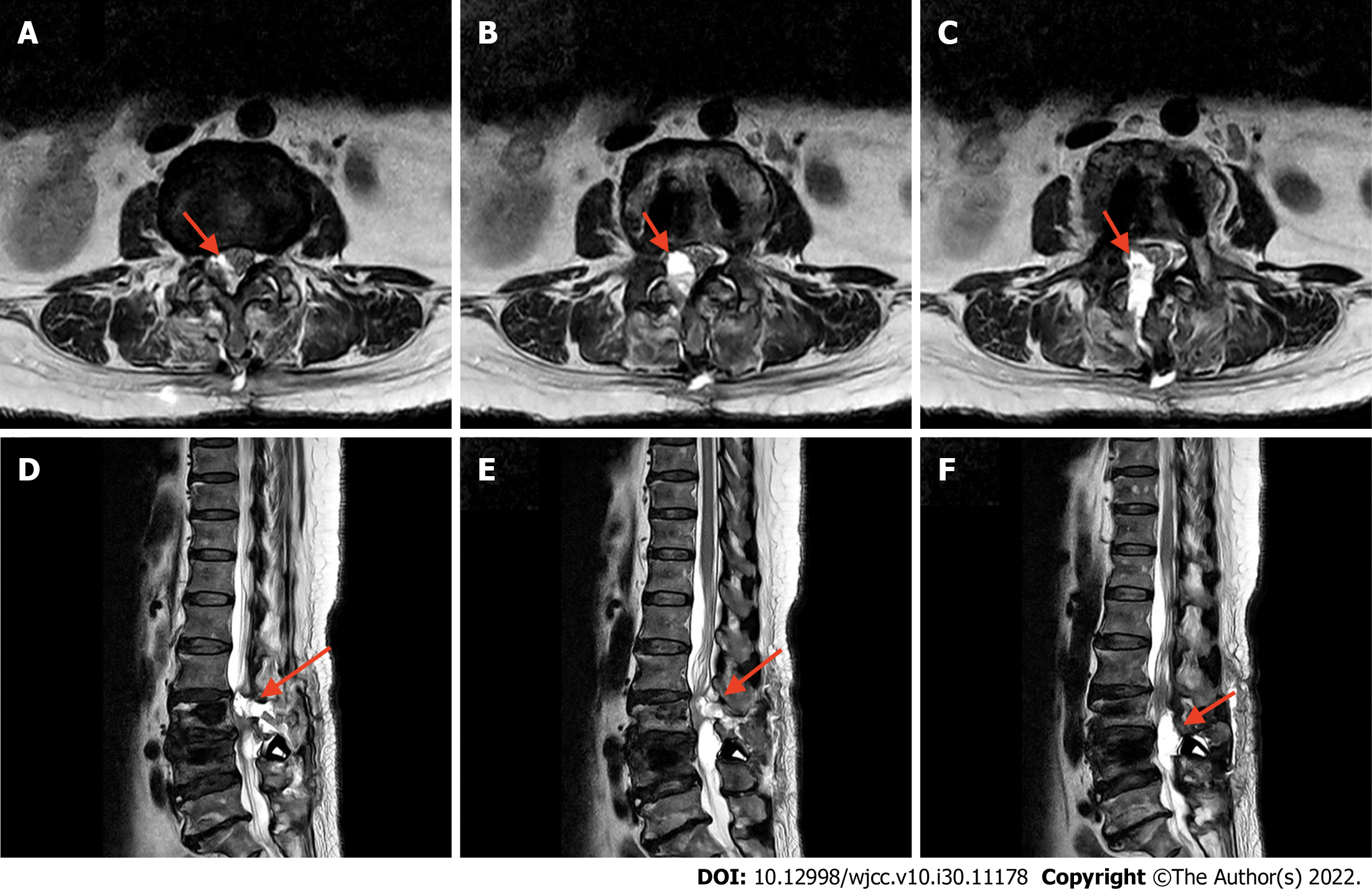
Figure 2 Postoperative T2-weighted magnetic resonance imaging.
A-C: Sagittal views; D-F: Axial views. Regional fluid collection at the surgical bed, protruding anteriorly at the junction of L2 and L3 toward the L4 Level are visible. The fluid has caused an indentation of the thecal sac and narrowing of the central canal.
Figure 3 Removal of the gel-like DuraSealTM.
The gel-like DuraSealTM is compressing the thecal sac and narrowing the central canal.
- Citation: Yeh KL, Wu SH, Fuh CS, Huang YH, Chen CS, Wu SS. Cauda equina syndrome caused by the application of DuraSealTM in a microlaminectomy surgery: A case report. World J Clin Cases 2022; 10(30): 11178-11184
- URL: https://www.wjgnet.com/2307-8960/full/v10/i30/11178.htm
- DOI: https://dx.doi.org/10.12998/wjcc.v10.i30.11178


