Copyright
©The Author(s) 2022.
World J Clin Cases. Oct 26, 2022; 10(30): 11139-11145
Published online Oct 26, 2022. doi: 10.12998/wjcc.v10.i30.11139
Published online Oct 26, 2022. doi: 10.12998/wjcc.v10.i30.11139
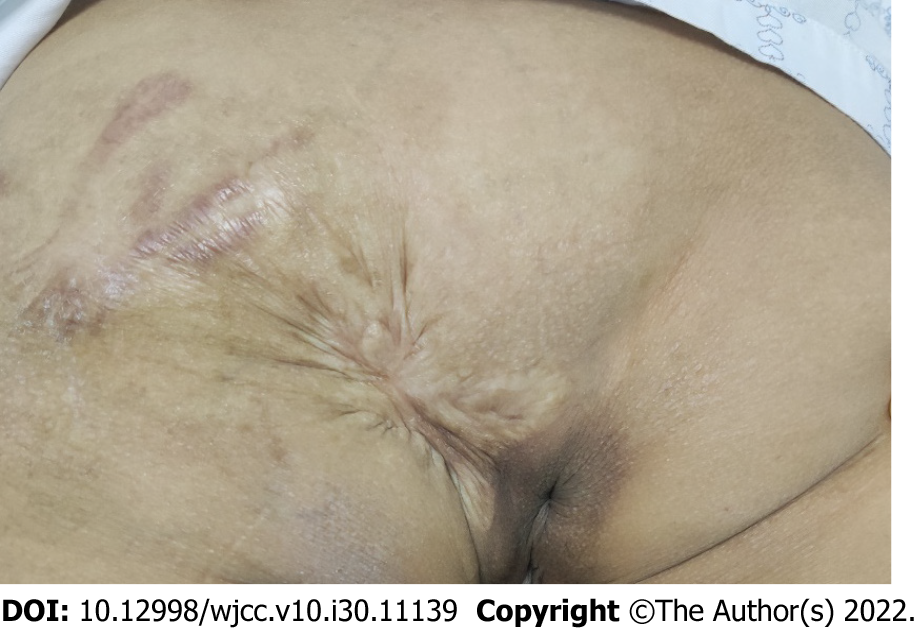
Figure 1 Preoperative image of the mass.
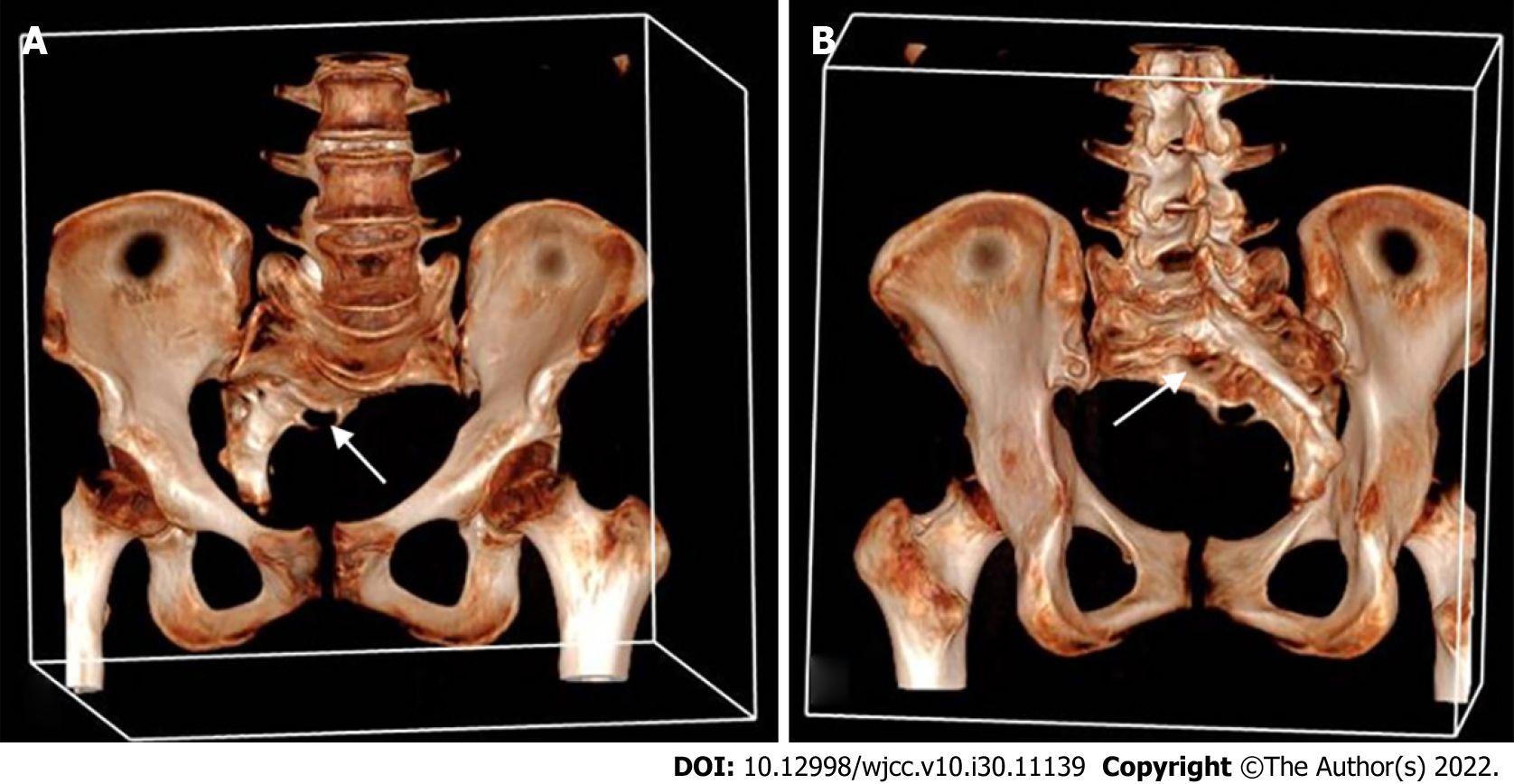
Figure 2 Computed tomography scan examination of the sacral vertebra.
A: Frontal view; B: Dorsal view: Three-dimensional computed tomography scan showed a sacrococcygeal scoliosis below the S2 level (white arrows); the sacral canal is partially enlarged and opened.
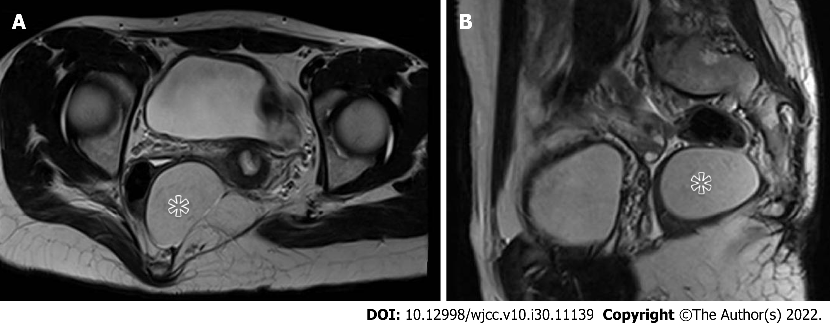
Figure 3 Magnetic resonance imaging.
A: T2-weighted imaging: a well-circumscribed mass (asterisk) compressing the rectum and displacing it right-anteriorly; B: T2-weighted imaging showed a well-defined mass anterior to the sacrum.
Figure 4 Axial T1-weighted imaging.
A: Circular signal (white arrows) on the outside of the levator ani muscle with strips signs connecting to the skin of the left buttock; B: Contrast-enhanced T1-weighted imaging showed enhancement of the strips signs.
- Citation: Ji ZX, Yan S, Gao XC, Lin LF, Li Q, Yao Q, Wang D. Perirectal epidermoid cyst in a patient with sacrococcygeal scoliosis and anal sinus: A case report. World J Clin Cases 2022; 10(30): 11139-11145
- URL: https://www.wjgnet.com/2307-8960/full/v10/i30/11139.htm
- DOI: https://dx.doi.org/10.12998/wjcc.v10.i30.11139


