Copyright
©The Author(s) 2022.
World J Clin Cases. Oct 26, 2022; 10(30): 10997-11003
Published online Oct 26, 2022. doi: 10.12998/wjcc.v10.i30.10997
Published online Oct 26, 2022. doi: 10.12998/wjcc.v10.i30.10997
Figure 1 Surgical image.
A: Skin rash on the patient’s back at presentation; B: Skin rash on the patient’s leg at presentation.
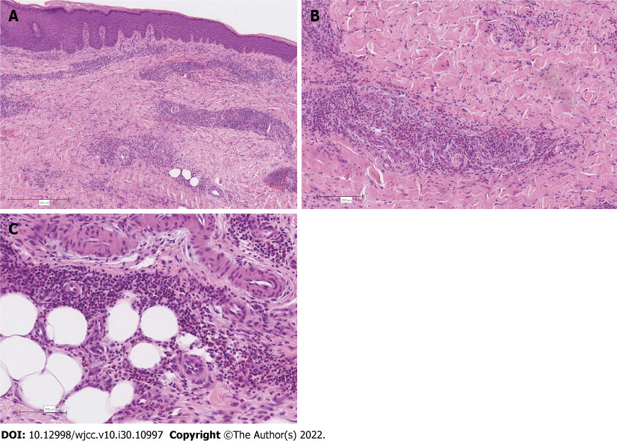
Figure 2 Histological evidence of Wells’ syndrome in skin from the right thigh.
A: Psoriasiform hyperplasia and moderately intense superficial and deep perivascular and interstitial inflammatory cell infiltrate are evident, extending into the subcutis (magnification: 40 ×); B: Eosinophilic granulocytes as the main component of the inflammatory cell infiltrate. Here, they are seen in perivascular areas but also within the interstitium between collagen bundles (magnification: 100 ×); C: Inflammatory cell infiltrate mainly composed of eosinophilic granulocytes extending into the subcutaneous fatty tissue (magnification: 200 ×).
- Citation: Šajn M, Luzar B, Zver S. Wells’ syndrome possibly caused by hematologic malignancy, influenza vaccination or ibrutinib: A case report. World J Clin Cases 2022; 10(30): 10997-11003
- URL: https://www.wjgnet.com/2307-8960/full/v10/i30/10997.htm
- DOI: https://dx.doi.org/10.12998/wjcc.v10.i30.10997


