Copyright
©The Author(s) 2022.
World J Clin Cases. Jan 21, 2022; 10(3): 1122-1130
Published online Jan 21, 2022. doi: 10.12998/wjcc.v10.i3.1122
Published online Jan 21, 2022. doi: 10.12998/wjcc.v10.i3.1122
Figure 1 Imaging documented the anal mass (see orange arrows).
A: Low-density shadows on the posterior edge of the anal canal in computed tomography; B and C: magnetic resonance imaging showed increased signal in anal tissue.
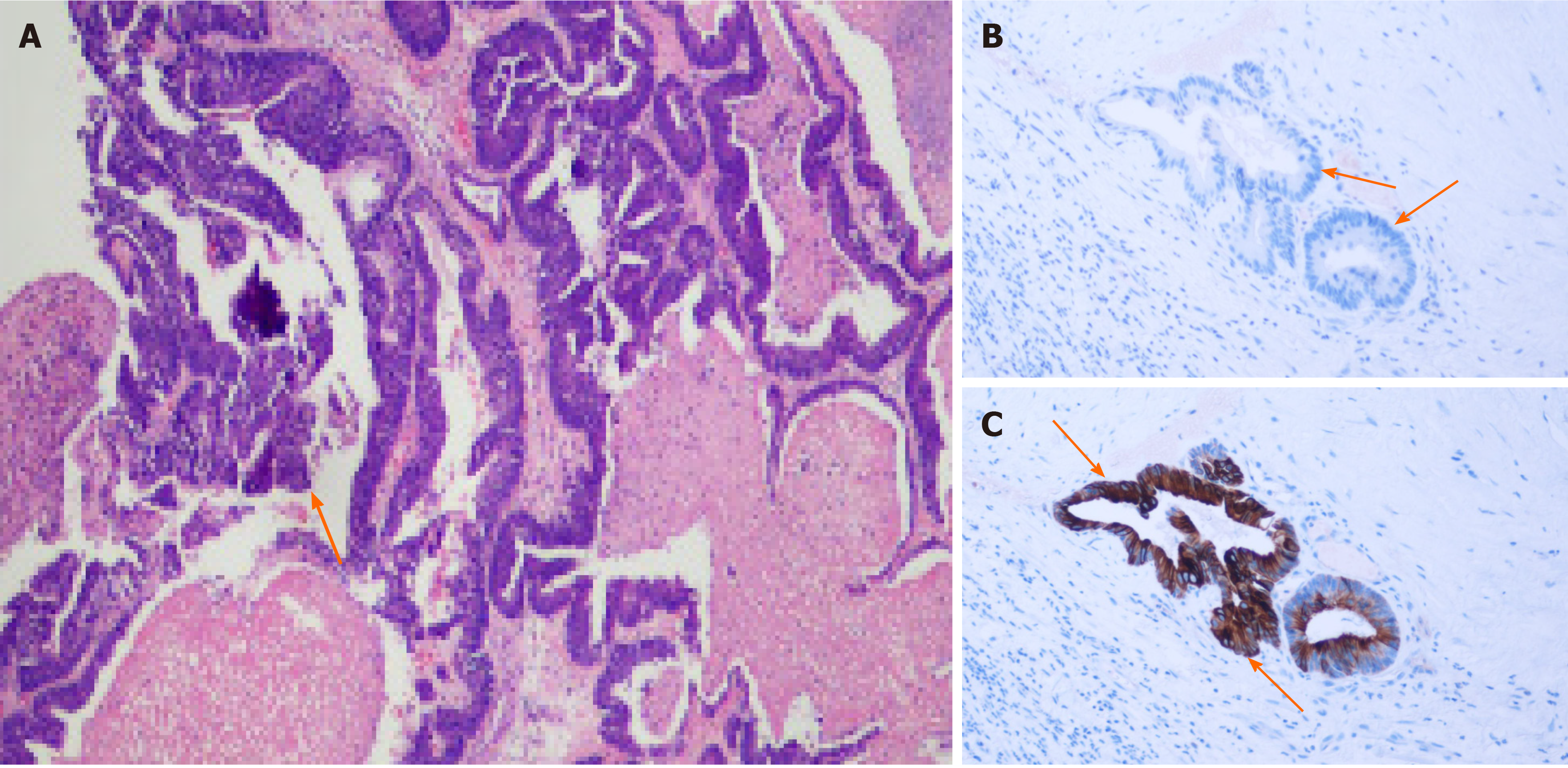
Figure 2 Pathology of anal mass.
A: Histology showed moderately differentiated adenocarcinoma, as orange arrow marked; B and C: Pathological staining for CK7 (B) and CK20 (C), with orange arrows marking negative and positive staining.
- Citation: Meng LK, Zhu D, Zhang Y, Fang Y, Liu WZ, Zhang XQ, Zhu Y. Recurrence of sigmoid colon cancer–derived anal metastasis: A case report and review of literature. World J Clin Cases 2022; 10(3): 1122-1130
- URL: https://www.wjgnet.com/2307-8960/full/v10/i3/1122.htm
- DOI: https://dx.doi.org/10.12998/wjcc.v10.i3.1122


