Copyright
©The Author(s) 2022.
World J Clin Cases. Oct 16, 2022; 10(29): 10811-10816
Published online Oct 16, 2022. doi: 10.12998/wjcc.v10.i29.10811
Published online Oct 16, 2022. doi: 10.12998/wjcc.v10.i29.10811
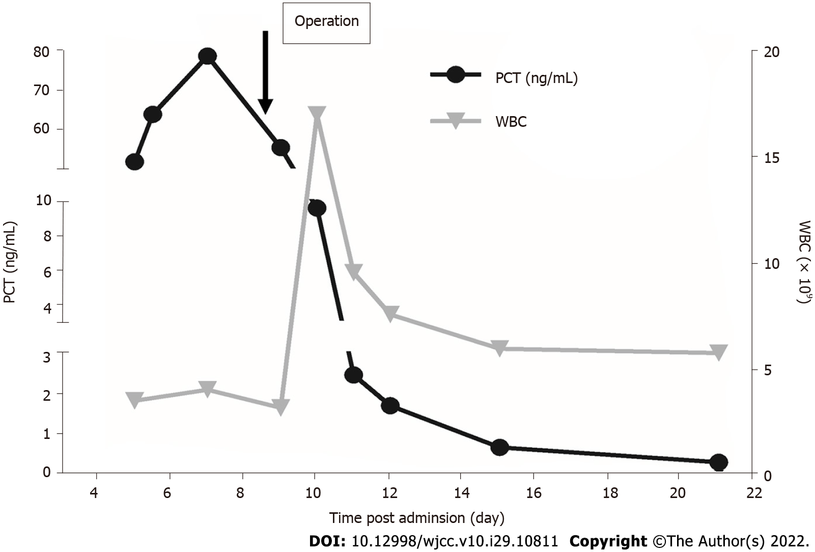
Figure 1 Dynamic changes in procalcitonin levels and count of white cells.
PCT: Procalcitonin; WBC: White blood cell.
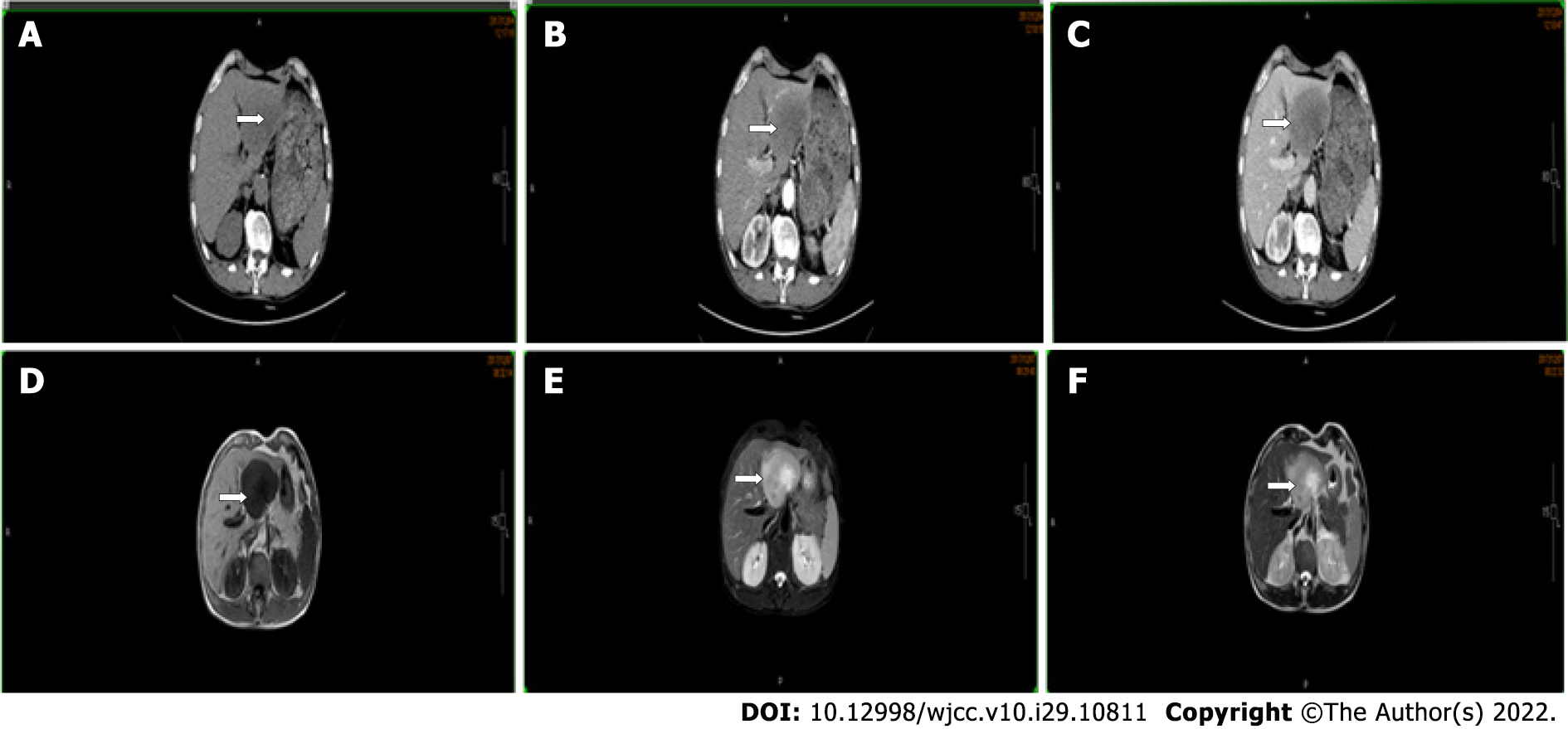
Figure 2 Abdominal computed tomography scan and magnetic resonance imaging show a tumor in the left lateral lobe (arrow).
A: No contrast phase; B: Hepatic arterial phase; C: Portal venous phase; D: T1; E: T2 weighted-turbo spin echo; F: T2.
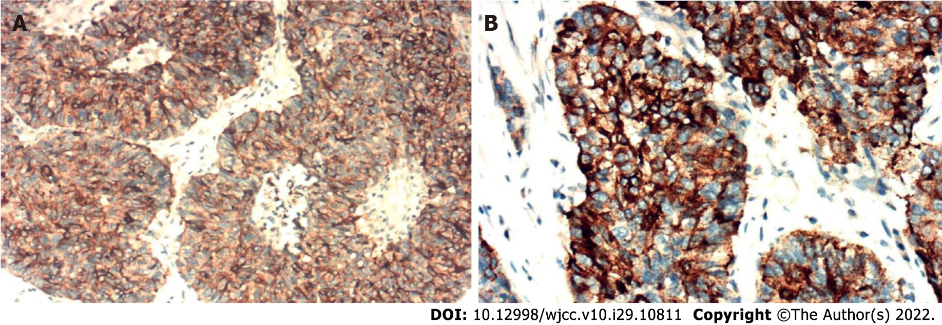
Figure 3 Immunohistochemical analyses revealed that tumor cells were positive for procalcitonin expression (A: 200 ×; B: 400 ×).
- Citation: Zeng JT, Wang Y, Wang Y, Luo ZH, Qing Z, Zhang Y, Zhang YL, Zhang JF, Li DW, Luo XZ. Elevated procalcitonin levels in the absence of infection in procalcitonin-secretin hepatocellular carcinoma: A case report. World J Clin Cases 2022; 10(29): 10811-10816
- URL: https://www.wjgnet.com/2307-8960/full/v10/i29/10811.htm
- DOI: https://dx.doi.org/10.12998/wjcc.v10.i29.10811


