Copyright
©The Author(s) 2022.
World J Clin Cases. Oct 16, 2022; 10(29): 10728-10734
Published online Oct 16, 2022. doi: 10.12998/wjcc.v10.i29.10728
Published online Oct 16, 2022. doi: 10.12998/wjcc.v10.i29.10728
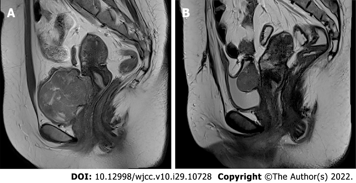
Figure 1 Magnetic resonance imaging findings.
A: Initial Magnetic resonance imaging (MRI) finding. Sagittal T2-weighted MR image shows a 6 cm × 4.6 cm mass between the uterine cervix and urinary bladder; B: MRI finding after recurrence. Sagittal T2-weighted MR image shows a 3.8 cm × 3 cm sized homogeneous mass arising from posterior wall of the bladder.
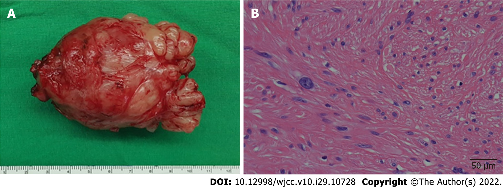
Figure 2 Initial pathological findings.
A: Gross image; B: Leiomyoma with bizarre nuclei and moderate cytologic atypia cells (hematoxylin-eosin stain, scale bar = 50 µm).
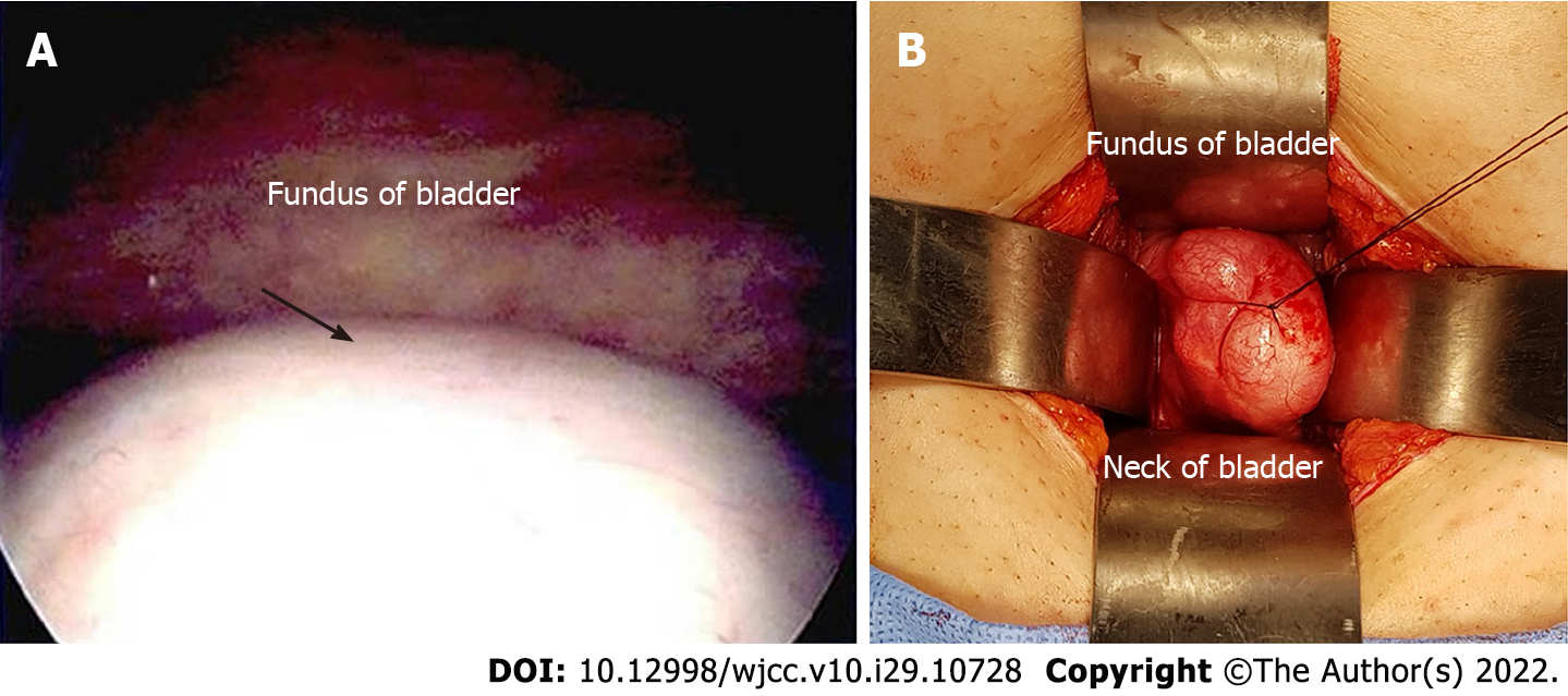
Figure 3 Cystoscopic and intraoperative findings after recurrence.
A: Cystoscopic image (arrow: Protruding mass); B: Intraoperative finding (protruding mass in the bladder).
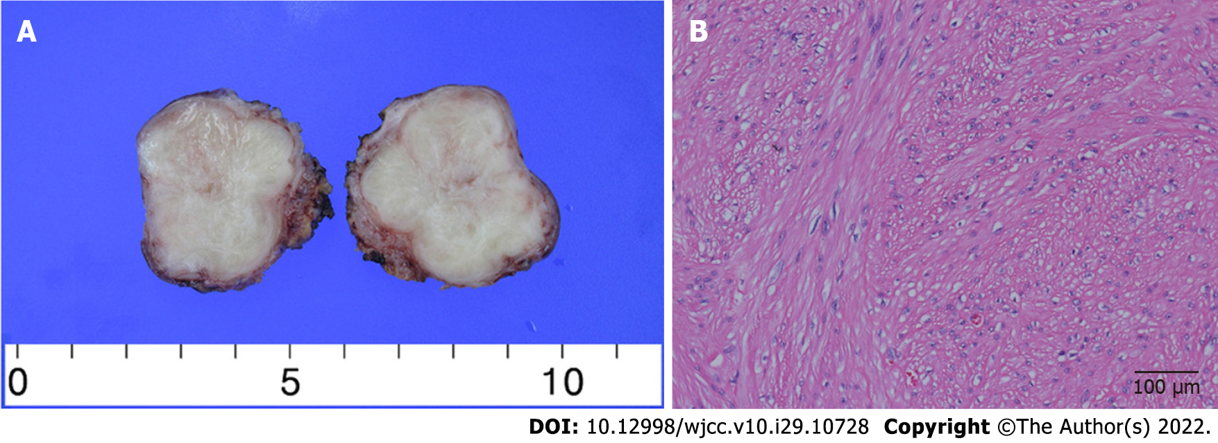
Figure 4 Pathological findings after recurrence.
A: Gross image; B: Leiomyoma without atypia (hematoxylin-eosin stain, scale bar = 100 µm).
- Citation: Song J, Song H, Kim YW. Recurrent atypical leiomyoma in bladder trigone, confused with uterine fibroids: A case report. World J Clin Cases 2022; 10(29): 10728-10734
- URL: https://www.wjgnet.com/2307-8960/full/v10/i29/10728.htm
- DOI: https://dx.doi.org/10.12998/wjcc.v10.i29.10728


