Copyright
©The Author(s) 2022.
World J Clin Cases. Oct 16, 2022; 10(29): 10655-10662
Published online Oct 16, 2022. doi: 10.12998/wjcc.v10.i29.10655
Published online Oct 16, 2022. doi: 10.12998/wjcc.v10.i29.10655
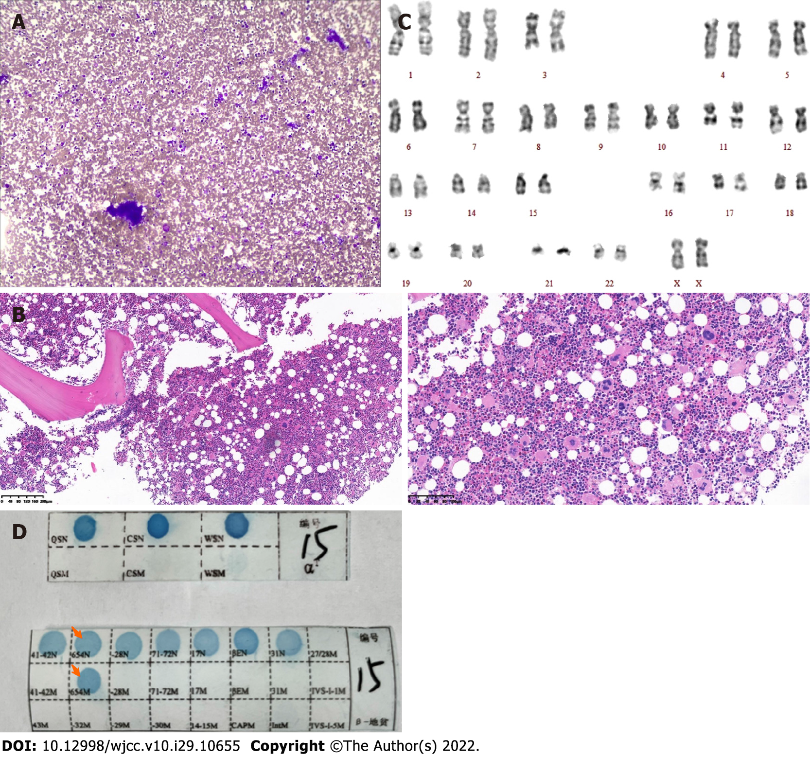
Figure 1 Histopathological changes, Karyotype and polymerase chain reaction and reverse dot blot observed in this case.
A: Hematoxylin and eosin staining; 100× bone marrow smear showing platelets in clusters; B: Hematoxylin and eosin staining of the trephine biopsy section (100×, 200×). The ratio of bone marrow hematopoietic tissue to fat cells was 80%:20%, and the granulocyte-to-erythrocyte ratio was approximately 1-2:1; granulocyte hyperplasia (+), erythroid hyperplasia (++), 25-30 megakaryocytes in each high-power field, a local clustering trend, pleomorphism, and reticular fibers (+) were all observed. Pathological diagnosis: right posterior superior spine bone marrow biopsy demonstrated chronic myeloproliferative neoplasms, characterized by megakaryocyte hyperplasia; C: Karyotype showing 46 chromosomes, [A1] including XX chromosomes (20 cells examined); D: PCR-based reverse dot blot showing β-IVS-Ⅱ-654-thalassemia.
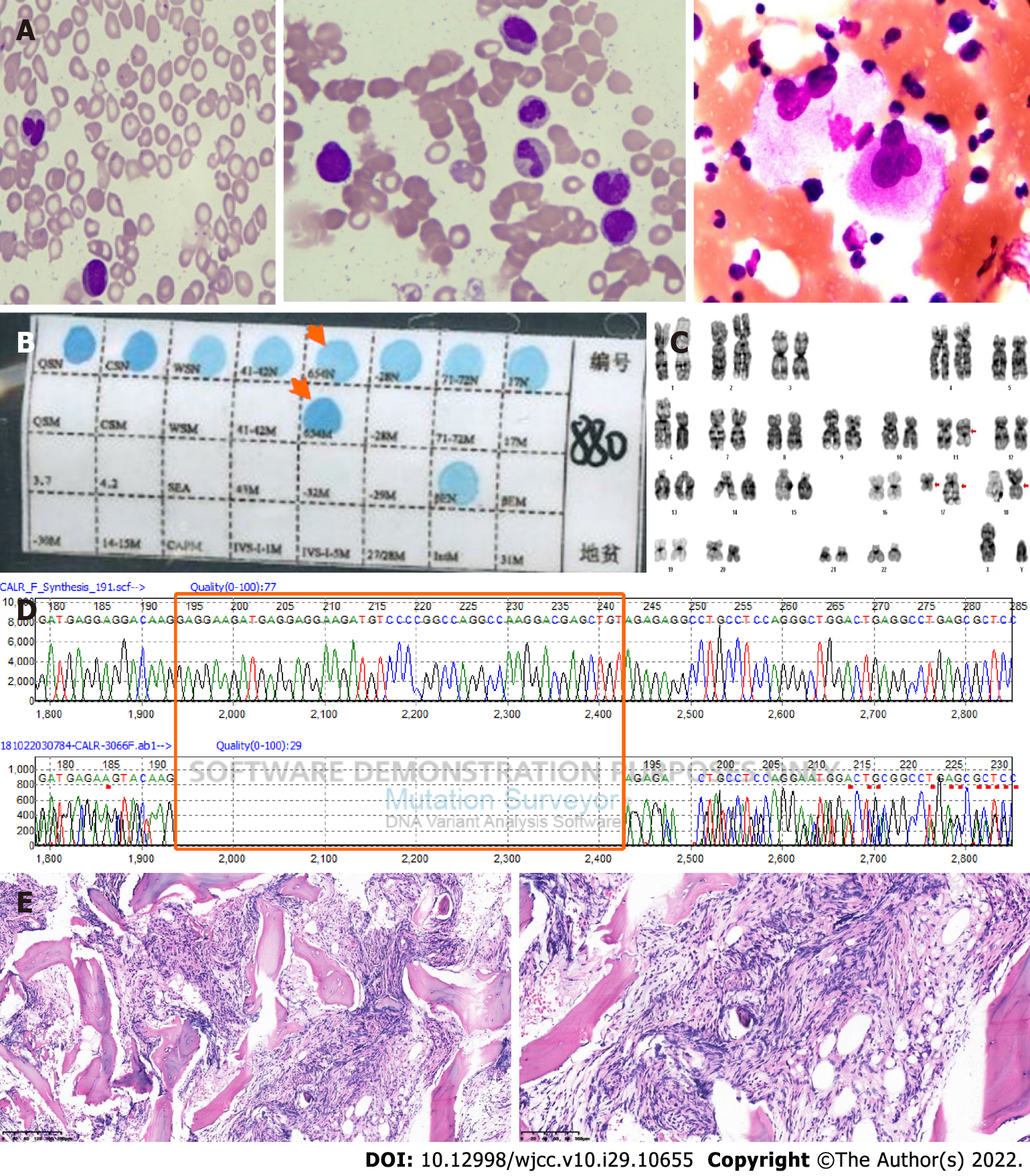
Figure 2 Bone marrow smear, biopsy section, Karyotype, Partial sequencing electropherogram and polymerase chain reaction and reverse dot blot observed in this case.
A: Hematoxylin and eosin staining (1000×, 100×) of the bone marrow aspirate showed teardrop-shaped, oval, long red blood cells, active hyperplasia of bone marrow granulocytes, erythroblastic cells that were hypoplastic, and a normal number of megakaryocytes with a decreased platelet production; B: PCR-based reverse dot blot showing β-IVS-II-654-thalassemia; C: Karyotype: 46 chromosomes, including XY, t(11; 18)(q13; p11.2), and ins(17; 17)(q21; q25q12)[20]; D: Partial sequencing electropherogram showing a type 1 (49-bp) deletion in exon 9 of CALR (194-242, GAG……. TGT); E: Hematoxylin and eosin staining (100×, 200×) showing BM reticulin (+++) with megakaryocyte hyperplasia and dysplasia.
- Citation: Xu NW, Li LJ. Myeloproliferative neoplasms complicated with β-thalassemia: Two case report. World J Clin Cases 2022; 10(29): 10655-10662
- URL: https://www.wjgnet.com/2307-8960/full/v10/i29/10655.htm
- DOI: https://dx.doi.org/10.12998/wjcc.v10.i29.10655


