Copyright
©The Author(s) 2022.
World J Clin Cases. Oct 16, 2022; 10(29): 10606-10613
Published online Oct 16, 2022. doi: 10.12998/wjcc.v10.i29.10606
Published online Oct 16, 2022. doi: 10.12998/wjcc.v10.i29.10606
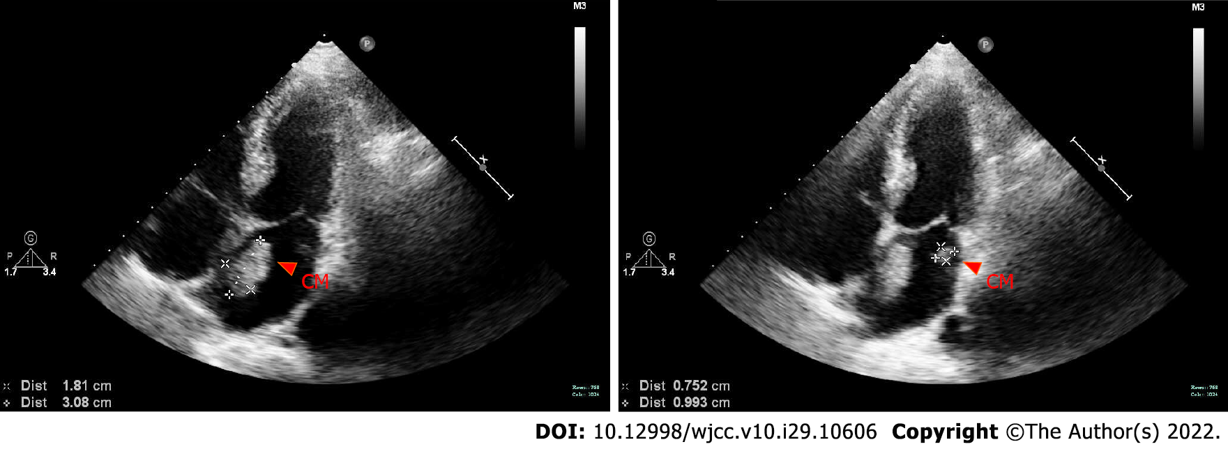
Figure 1 Cardiac ultrasound.
Two abnormal psychogenic masses were seen in the left atrium, 3.1 cm × 1.8cm and 1.0 cm × 0.8cm respectively. CM: Cardiac myxoma.
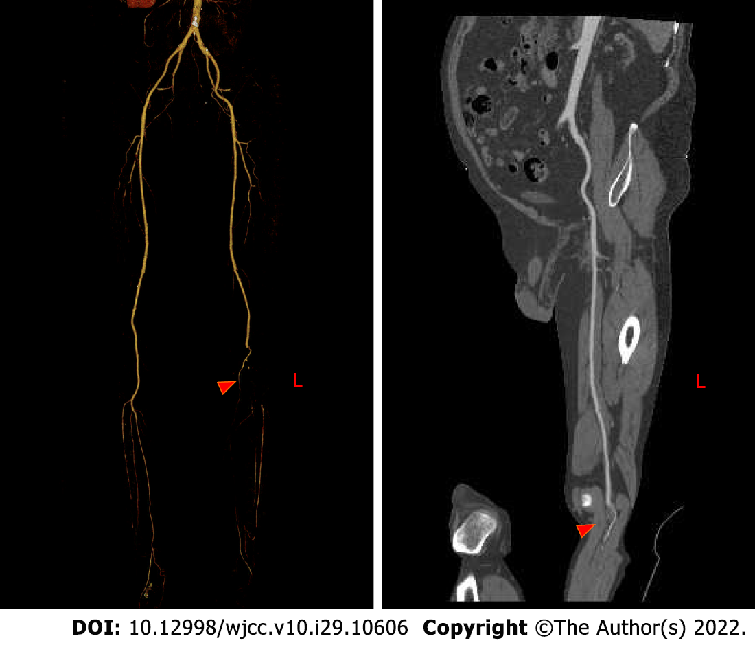
Figure 2 Computer tomography hagiography of lower extremity blood vessels before operation.
The left political artery was not visualized, the lumen embolism was considered, the peritoneal artery and the posterior tibial artery were occluded, and the proximal end of the anterior tibial artery was occluded.
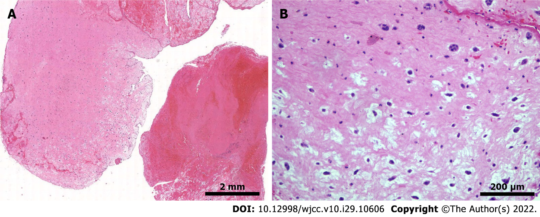
Figure 3 Hematological staining.
A: Histologist section shows embolism with thrombus components and mucous background, with scattered sparse cell clusters and multiple villi-like protrusions, which are consistent with the characteristics of soft and brittle taxonomy (1 ×); B: High magnification microscope shows scattered free clusters on a mucus background with nested round, polygonal cells (squamous cells) arranged in short cords, with round nuclei and fine chromatin, and no atypical (10 ×).
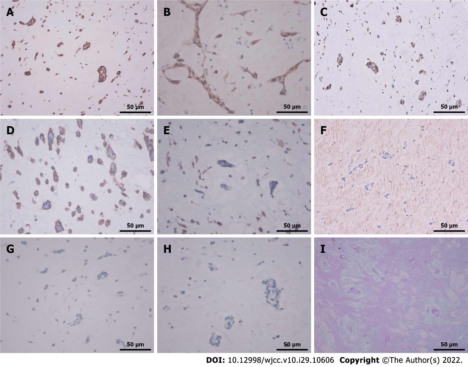
Figure 4 Immunodeficiency, alliance blue staining and periodic acid skiff staining (40 ×).
A: Cytoplasmic sentiment (sentiment) positive; B: Cytoplasmic and nuclear retinal positive; C: Cytoplasmic CD31 positive; D: Cytoplasmic CD34 positive; E: Cytoplasmic CD68 negative; F: Cytoplasmic Factor 8 related antigen negative; G: Cytoplasmic cytokines protein was negative; H: Cytoplasmic cytokines 5/6 was negative; I: Cytoplasmic alliance blue staining and periodic acid skiff staining was positive.
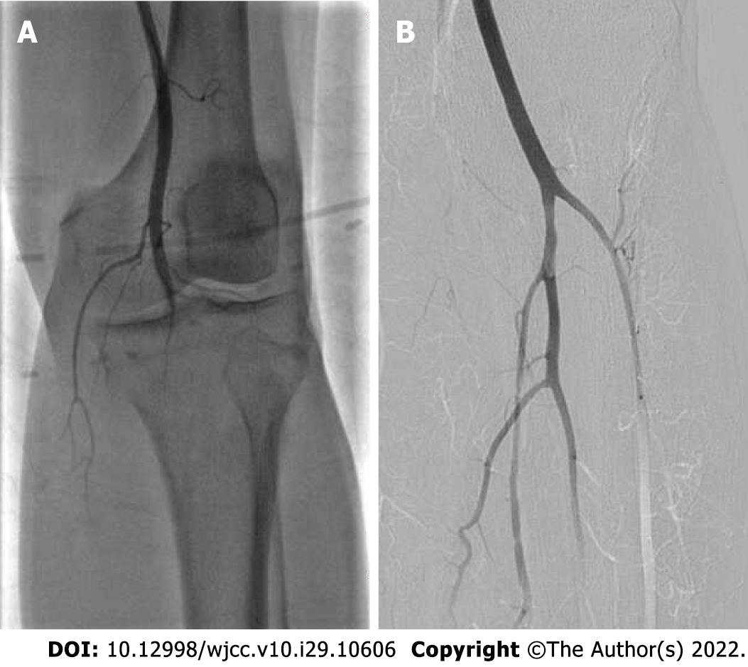
Figure 5 Angiography results.
A: Lower extremity historiography before thrombolytic showed that the political artery was occluded, and the infra-knee artery was not visualized; B: Lower extremity historiography after thrombolytic showed that the blood flow of the political artery, tribulation trunk, anterior tibial artery and peritoneal artery was unobstructed.
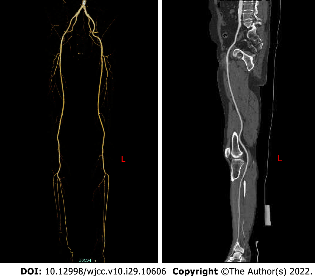
Figure 6 Postoperative re-examination of lower extremity vascular computer tomography angiography showed that the left political artery, anterior tibial artery and peritoneal artery were still unobstructed.
- Citation: Meng XH, Xie LS, Xie XP, Liu YC, Huang CP, Wang LJ, Zhang GH, Xu D, Cai XC, Fang X. Cardiac myxoma shedding leads to lower extremity arterial embolism: A case report. World J Clin Cases 2022; 10(29): 10606-10613
- URL: https://www.wjgnet.com/2307-8960/full/v10/i29/10606.htm
- DOI: https://dx.doi.org/10.12998/wjcc.v10.i29.10606


