Copyright
©The Author(s) 2022.
World J Clin Cases. Oct 16, 2022; 10(29): 10565-10574
Published online Oct 16, 2022. doi: 10.12998/wjcc.v10.i29.10565
Published online Oct 16, 2022. doi: 10.12998/wjcc.v10.i29.10565
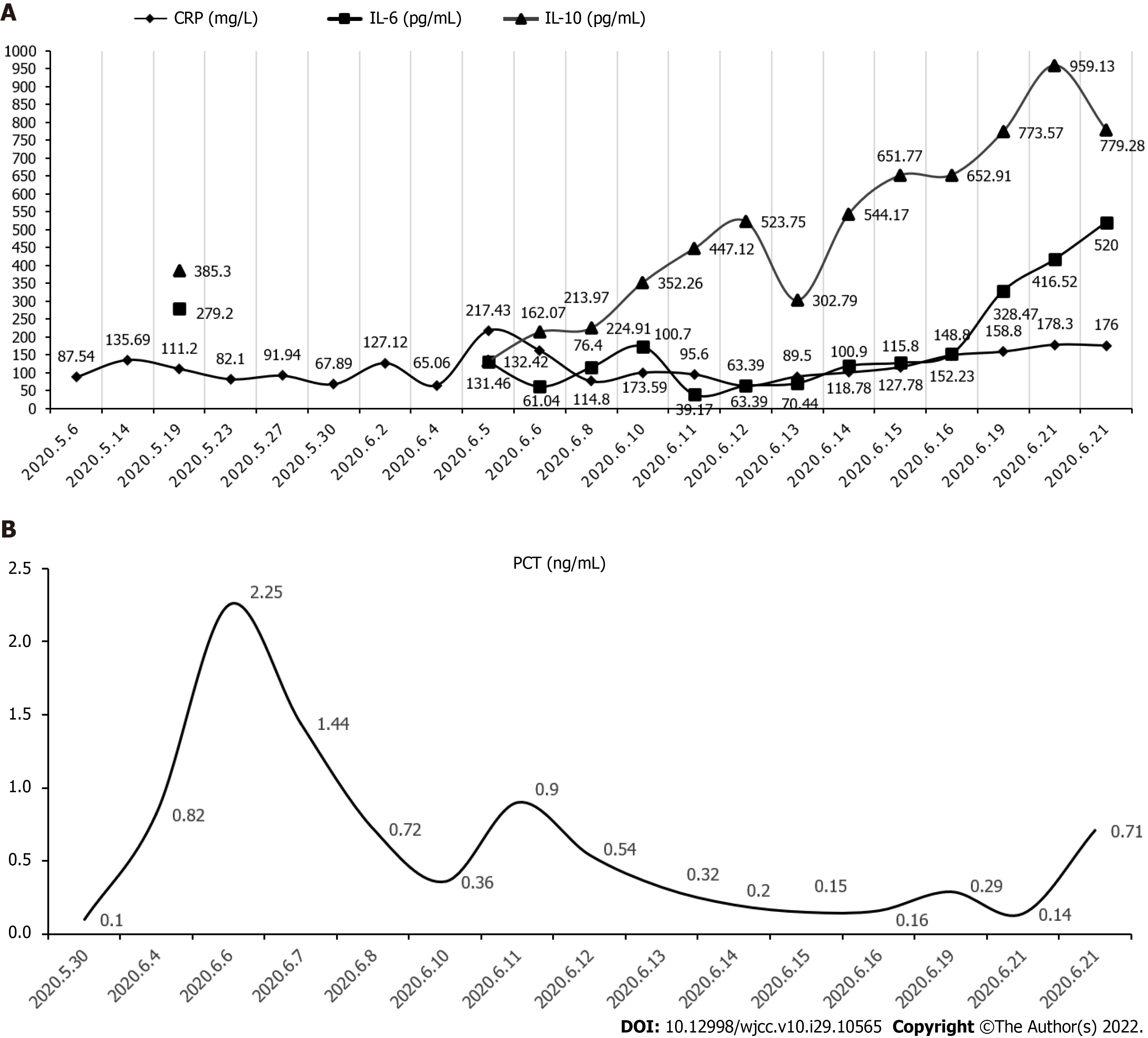
Figure 1 The potential changes in inflammatory biomarkers of this patient.
A: The observed changes in C-reactive protein, interleukin (IL)-10, and IL-6 levels; B: The changes of procalcitonin levels. CRP: C-reactive protein; IL: Interleukin; PCT: Procalcitonin.
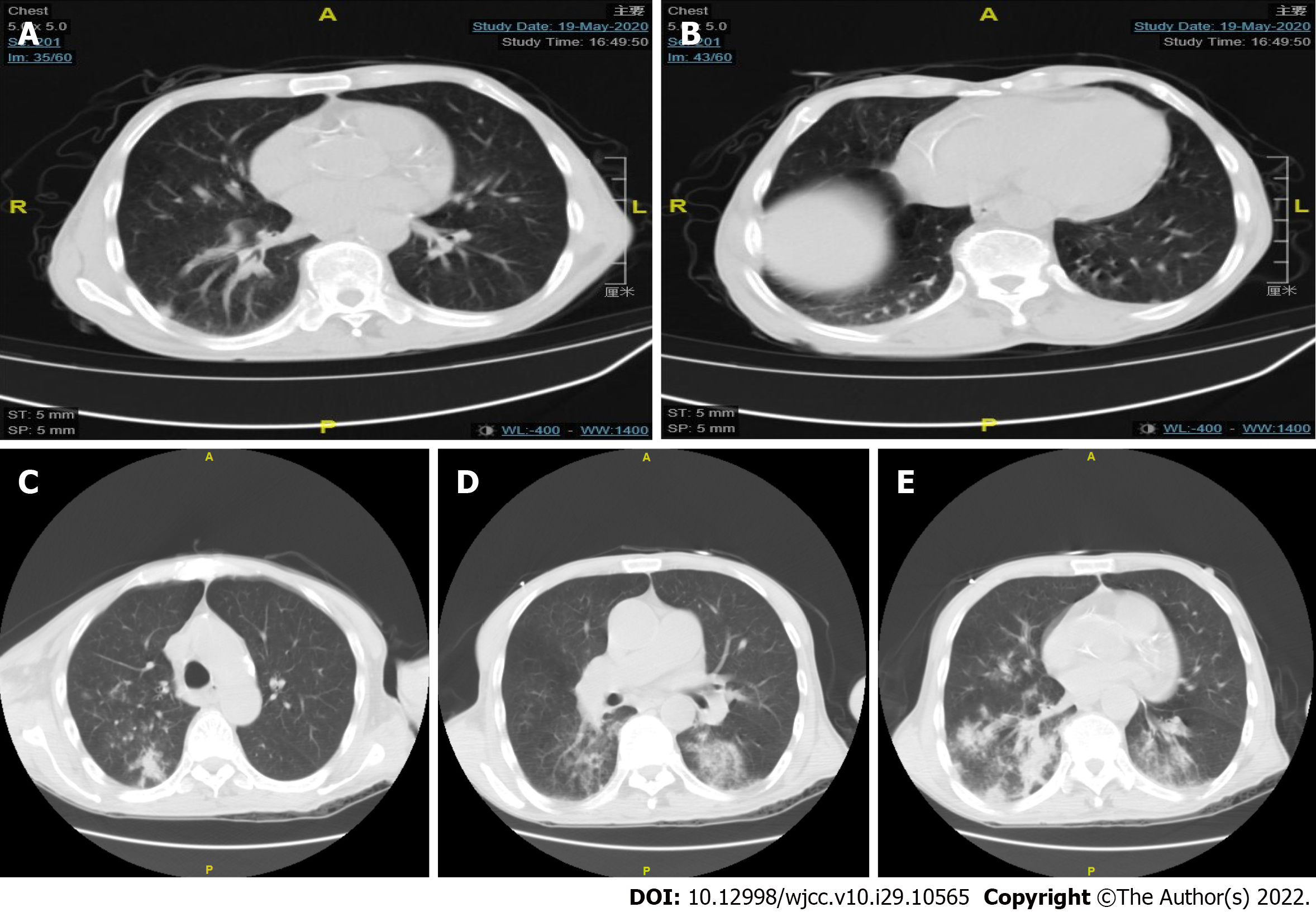
Figure 2 Chest computed tomography findings of the patient in two hospitalizations.
A and B: The patient's first chest computed tomography (CT) after the onset of illness. As shown by the arrows, a small amount of pleural effusion can be seen in both the lungs, and scattered nodules can be observed in the pleura and under the pleura; C-E: The results of chest CT upon two weeks later. The new appearances of the two lungs were mostly scattered in the patchy high-density shadows, with distinctly blurred boundaries, distributed along with the bronchial vascular bundle, mainly under the pleura.
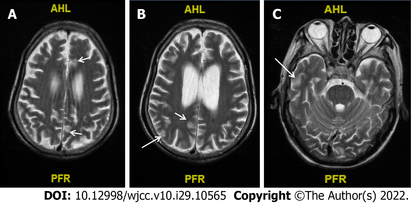
Figure 3 The result of cranial magnetic resonance upon the patient's second admission.
A-C: Abnormal signals diverged from the subfrontal cortex (A), midbrain (B), and the posterior horns of both sides of the ventricle (C) (arrow pointing), and no obvious brain abscess formation was noted.
- Citation: Wu GX, Zhou JY, Hong WJ, Huang J, Yan SQ. Treatment failure in a patient infected with Listeria sepsis combined with latent meningitis: A case report. World J Clin Cases 2022; 10(29): 10565-10574
- URL: https://www.wjgnet.com/2307-8960/full/v10/i29/10565.htm
- DOI: https://dx.doi.org/10.12998/wjcc.v10.i29.10565


