Copyright
©The Author(s) 2022.
World J Clin Cases. Oct 16, 2022; 10(29): 10559-10564
Published online Oct 16, 2022. doi: 10.12998/wjcc.v10.i29.10559
Published online Oct 16, 2022. doi: 10.12998/wjcc.v10.i29.10559
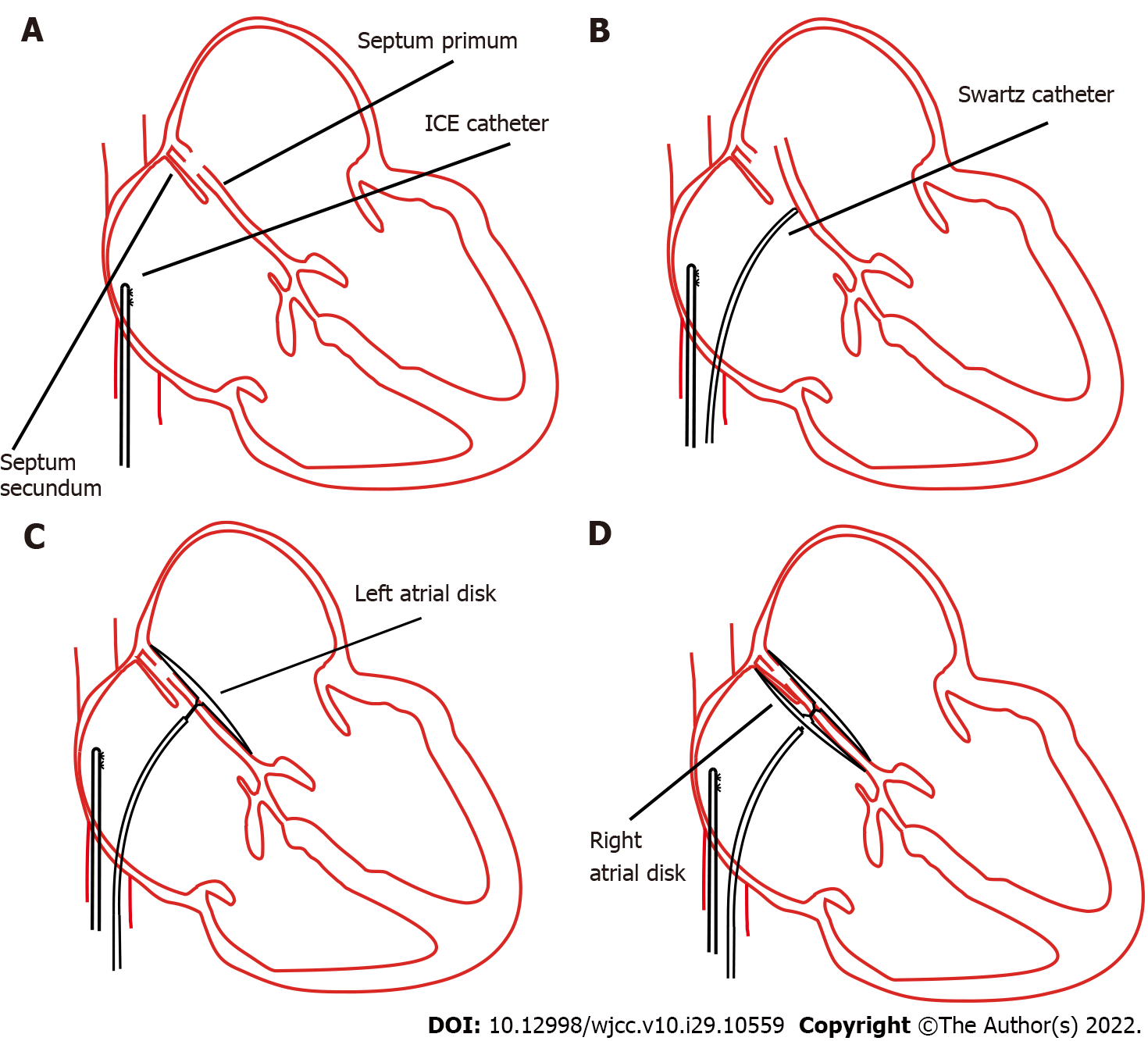
Figure 1 Concept procedures for percutaneous patent foramen ovale closure through intracardiac echocardiography guidance.
A: The intracardiac echocardiography catheter was advanced into the right atrium; B: The septum primum was artificially separated from the septum secundum by applying physical pressure using a Swartz sheath; C: Deployment of the right atrial disk; D: Complete deployment of right and left side disks. ICE: Intracardiac echocardiography.
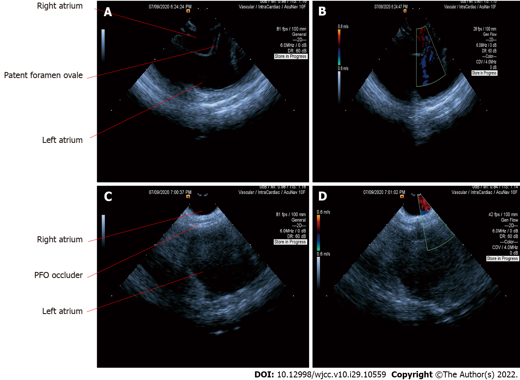
Figure 2 Intracardiac echocardiography images in the diagnosis and closure of patent foramen ovale.
A: Separation of the septum primum and septum secundum through a Swartz sheath; B: Patent foramen ovale (PFO) was confirmed using a colour Doppler; C: The septum primum and septum secundum were fixed together after complete deployment of right and left side disks; D: Confirmation of PFO closure using a colour Doppler. PFO: Patent foramen ovale.
- Citation: Han KN, Yang SW, Zhou YJ. Novel way of patent foramen ovale detection and percutaneous closure by intracardiac echocardiography: A case report. World J Clin Cases 2022; 10(29): 10559-10564
- URL: https://www.wjgnet.com/2307-8960/full/v10/i29/10559.htm
- DOI: https://dx.doi.org/10.12998/wjcc.v10.i29.10559


