Copyright
©The Author(s) 2022.
World J Clin Cases. Oct 6, 2022; 10(28): 10266-10272
Published online Oct 6, 2022. doi: 10.12998/wjcc.v10.i28.10266
Published online Oct 6, 2022. doi: 10.12998/wjcc.v10.i28.10266
Figure 1 Coronary artery computed tomography angiography of the Kawasaki disease patient.
A: Multiple coronary artery aneurysms, and multiple thrombi in the coronary artery ectasia of the proximal segment of the right coronary artery. B: The ectatic coronary artery were observed in the extremity of left main coronary artery. C: Two coronary artery aneurysms with vascular calcification were observed in the left circumflex artery.
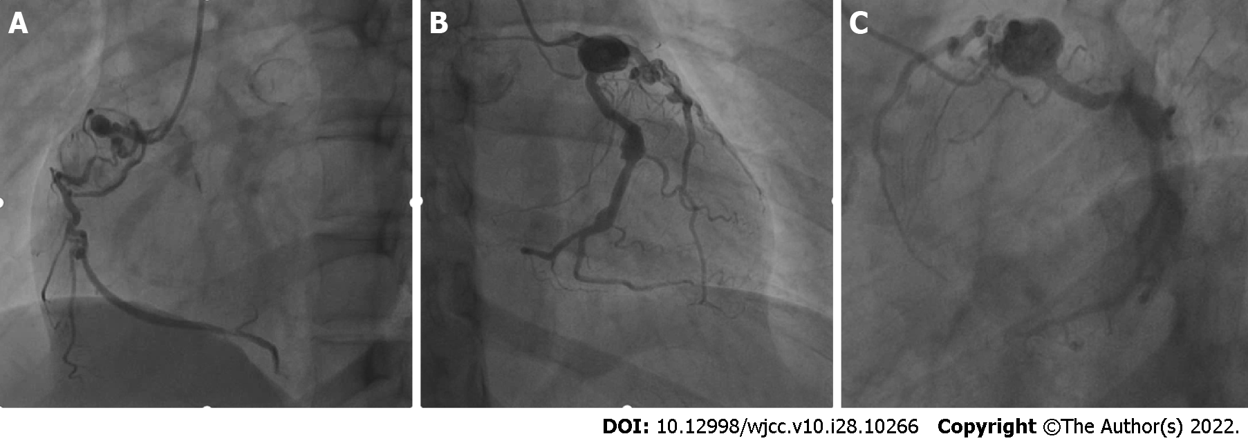
Figure 2 Coronary angiography of the Kawasaki disease patient.
A: Big coronary artery aneurysm was in a proximal segment of the right coronary artery with an organized thrombus; B: Two hemangiomas were observed at the extremity of the left circumflex artery with calcification; C: The vessels in the descending proximal left anterior were tortuous with thrombus, and the distal vessels were in the myocardial bridge.
Figure 3 Doppler echocardiography of the Kawasaki disease patient.
A: The inner diameter of the left main coronary artery was 0.4 cm; B: The inner diameter of the aneurysm near the cross of vessels was 1.0 cm; C: The inner diameter of the right coronary artery was 0.56 cm.
- Citation: He Y, Ji H, Xie JC, Zhou L. Coronary artery aneurysms caused by Kawasaki disease in an adult: A case report and literature review. World J Clin Cases 2022; 10(28): 10266-10272
- URL: https://www.wjgnet.com/2307-8960/full/v10/i28/10266.htm
- DOI: https://dx.doi.org/10.12998/wjcc.v10.i28.10266


